Most people who come to an oculoplastic surgeon about heavy, hooded upper eyelids have never heard the word dermatochalasis. Their primary care doctor may have called it "droopy eyelids." A spa aesthetician may have suggested laser resurfacing. The condition is one of the most common reasons for upper eyelid surgery, yet patients almost never encounter its name before sitting down for a consultation. The gap matters, because dermatochalasis is not the same as a low lid margin, and it is not the same as a descending brow - and treating one when another is the actual problem produces results that disappoint or, in some cases, require revision. This page explains what dermatochalasis is at the tissue level, what happens to the anatomy as the condition develops, how a trained surgeon distinguishes it from the two conditions it most closely resembles, and what the clinical examination actually measures.
What Dermatochalasis Means
Dermatochalasis is the medical term for excess, redundant upper eyelid skin. The word comes from Greek: dermato meaning skin, and chalasis meaning relaxation or slackening. In plain terms, it describes a state in which the upper eyelid skin has lost enough structural integrity that it drapes downward over the lid margin, partially or fully obscuring the natural eyelid crease and, in more advanced cases, encroaching on the upper visual field.

The Anatomy That Changes
The upper eyelid is built in layers. From the surface inward: skin, a thin subcutaneous plane, the orbicularis oculi muscle (the sphincter-like muscle that closes the eye), the orbital septum, the preaponeurotic fat compartments, the levator aponeurosis, and the tarsus - the stiff cartilage-like plate that gives the lid its form. Dermatochalasis primarily affects the outermost layers, but the changes extend deeper than skin alone.
The Orbital Septum
The orbital septum is a fibrous membrane running from the periosteum of the orbital rim down to insert on the levator aponeurosis just above the top of the tarsal plate. Its job is to act as a barrier - holding orbital fat behind the lid and preventing it from pushing forward. With age, the septum progressively weakens and thins. Once it loses tensile strength, it can no longer contain the fat compartments that sit directly behind it.
The Two Fat Compartments
This is the portion of upper eyelid anatomy that most patient-facing resources omit entirely, even though it directly explains the fullness and heaviness that often accompanies hooded skin. The upper eyelid contains two distinct fat compartments, separated from each other by a thin interpad septum:
- The nasal (medial) fat pad - positioned toward the inner corner of the eye, this compartment contains white, fibrous fat. It is paler and firmer than the adjacent compartment.
- The central preaponeurotic fat pad - positioned toward the center of the lid, this compartment contains yellow, softer fat that sits directly in front of the levator aponeurosis.
As the orbital septum weakens, fat from either compartment - or both - can herniate forward through the lid, creating a visible bulge that adds to the appearance of hooding over and above the excess skin. A surgeon evaluating the upper lid assesses both compartments independently, because the degree of fat prolapse varies between patients and between the two sides of the same face. Removing skin without addressing herniated fat, when fat herniation is present, leaves the lid looking full and heavy even after the skin excess is corrected.
How Aging and Genetics Drive the Condition
The structural changes in dermatochalasis are not simply a matter of skin becoming loosely stretched. Histopathological analysis - microscopic examination of surgically removed eyelid tissue - reveals specific and consistent abnormalities in the tissue itself. Analysis of dermatochalasis specimens consistently shows three findings:
- Epidermal thinning and atrophy - the outer skin layer becomes thinner and loses its normal cellular architecture
- Disorganization and loss of dermal collagen - the collagen fibers that give skin tensile strength become widely spaced and atrophic rather than running in organized arrays
- Marked decrease and fragmentation of elastic fibers throughout the dermis - the elastic network that allows skin to recoil after deformation becomes sparse and broken
A 2011 study in Ophthalmology - the journal of the American Academy of Ophthalmology - identified elastolysis (enzymatic breakdown of elastic fibers) and lymphostasis as the fundamental, interrelated findings. The lymphostasis component is significant: disrupted lymphatic drainage in the eyelid tissue appears to contribute to structural deterioration alongside the direct loss of elastic and collagen proteins. A 2024 study published in Scientific Reports added another layer, finding that the integrity of the orbicularis oculi muscle also influences how dermatochalasis develops. The condition is not purely passive skin aging - there is an interplay between the sphincter muscle beneath the skin and the overlying connective tissue.
Why the Fifth Decade Is When Most People Notice It
Elastin production remains relatively steady through roughly the first four to five decades of life. After that point, production drops significantly. This partly explains the clinical pattern surgeons see routinely: patients who had no visible problem through their 30s and then noticed a rapid change in their mid-40s to early 50s. The decline in elastin synthesis is not sudden, but the threshold at which remaining elastic tissue can no longer compensate for ongoing breakdown shifts, and the accumulation becomes visible. Cumulative ultraviolet exposure accelerates this timeline - UV radiation degrades dermal collagen and elastic fibers faster than intrinsic aging alone.
Genetic and Other Risk Factors
Genetic predisposition is the single strongest risk factor for dermatochalasis. Familial patterns are common and clinically striking - some patients with a strong family history develop significant eyelid hooding in their 20s and 30s, two to three decades before typical age-related onset. Additional independent risk factors documented in the oculoplastic literature include:
- Higher BMI
- Male sex
- Lighter skin pigmentation
- Active smoking

Three Conditions That Look Alike but Are Not the Same
A heavy, tired, or visually obstructed upper eyelid region can result from three distinct anatomical sources, each requiring a different surgical correction. Treating one when another is the actual diagnosis produces a poor result - and in some combinations, an overcorrection or undercorrection that is difficult to revise.
| Condition | Anatomical Source | Lid Margin Height | Levator Function | Correct Fix |
|---|---|---|---|---|
| Dermatochalasis | Excess skin draping over lid; possible fat herniation | Normal | Intact and full | Upper blepharoplasty |
| True ptosis | Levator aponeurosis dehiscence or levator muscle weakness | Low | Reduced or poor | Ptosis repair (levator advancement or suspension) |
| Brow ptosis | Brow descending below the orbital rim | May appear low | Normal | Brow lift (direct, endoscopic, or transblepharoplasty approach) |
It is also worth distinguishing dermatochalasis from a condition with a similar-sounding name: blepharochalasis. Blepharochalasis is a rare inflammatory disorder marked by recurrent attacks of painless eyelid edema, typically in young people. Over years, these episodes cause skin atrophy, fat atrophy, and stretching of the levator aponeurosis - producing thin, fragile eyelid skin rather than the thickened, redundant skin of age-related dermatochalasis. The names cause confusion even among non-specialists, but the histology, patient age, clinical course, and management are entirely different.
Pseudoptosis: When Heavy Skin Fakes a Low Lid Margin
In most cases of dermatochalasis, the lid margin sits at its correct anatomical height. The skin is excessive; the lid position is not low. But in advanced cases, the weight of accumulated skin becomes sufficient to physically press the lid margin downward. On casual inspection - a photograph, a brief mirror check, a glance from a non-specialist - this looks indistinguishable from true ptosis. It is called pseudoptosis, because the lid margin is not genuinely low; it is being pushed down by the tissue above it.
The clinical test is direct: the examiner gently lifts the overhanging skin off the lid margin using a cotton-tipped applicator or a fingertip and reassesses lid position with the skin held clear. When the margin returns to a normal height once the skin is removed from the picture, the finding is pseudoptosis from dermatochalasis. When the margin stays low even after the skin is lifted, concurrent true ptosis is present and must be addressed as a separate problem.
The Clinical Examination: What Each Measurement Means
An oculoplastic evaluation for upper lid heaviness is a sequence of specific tests, not a general impression. Understanding what each one measures makes the consultation more informative and helps patients understand what the surgeon is actually determining. A complete exam moves through these steps in order:
- External inspection - assessing skin crease height, the extent of skin overhang, and whether the brow position appears to be contributing
- MRD1 measurement - establishing whether the lid margin sits at its correct anatomical height
- Pseudoptosis check - manually lifting excess skin to test whether the lid margin is being held down by skin weight
- Levator function measurement - documenting the full travel of the lid margin from maximal downgaze to maximal upgaze with the brow held firm
- Manual brow elevation test - isolating true lid skin excess from the contribution of brow descent
Marginal Reflex Distance 1
The surgeon shines a small light at the patient's eye while the patient looks straight ahead. The distance from the corneal light reflex to the upper lid margin is the marginal reflex distance 1, or MRD1. The commonly cited normal range is approximately 3-5 mm, with 4-5 mm used as a practical clinical reference in many blepharoplasty contexts - confirm the current reference range with your surgeon or an AAO resource, as values at the lower end of normal exist in some individuals. In uncomplicated dermatochalasis, MRD1 is normal - the levator muscle is lifting the lid to its correct height, and the excess skin is simply draping over it. In true ptosis, MRD1 falls below normal, reflecting a lid margin that is genuinely too low regardless of what the overlying skin is doing.
Skin Crease Height
The upper eyelid skin crease - the fold where the levator aponeurosis attaches to the skin - normally sits approximately 7-9 mm above the lid margin in men and 8-11 mm in women (these figures apply to Caucasian anatomy; the crease is typically lower or less defined in East Asian anatomy, which has distinct structural differences in fat distribution). In dermatochalasis, redundant skin descends below this crease and can reach the lash line, which produces the visual appearance of hooding. Documenting how far the skin has descended informs how much tissue needs to be excised and where the incision should be placed.
Levator Function
Levator function is the total travel of the upper lid margin, measured in millimeters, from maximal downgaze to maximal upgaze - while the examiner holds the brow firmly against any compensatory movement. Normal levator function is generally cited as 15 mm or more in the oculoplastic literature, with classification systems varying in their precise thresholds; values below about 12 mm are clearly reduced and values below 4 mm represent poor function. Dermatochalasis leaves levator function entirely intact; the muscle and its aponeurosis are not part of the problem. If excursion falls below the normal range, the levator aponeurosis may have weakened or partially dehisced, and blepharoplasty alone will not correct the underlying mechanism. That is precisely why surgeons test levator function during the exam even when the main complaint is about skin.
The Manual Brow Elevation Test
The examiner places a finger at the brow and holds it at the anatomically correct height - just above or at the orbital rim - while the patient fully relaxes the forehead. With the brow held in position, the surgeon reassesses how much eyelid skin remains redundant. If a significant portion of the apparent excess disappears when the brow is elevated, a meaningful contribution from brow ptosis is present. A patient in that situation who undergoes blepharoplasty without addressing the brow will be under-corrected, because skin that appeared to belong to the lid was actually being pushed there by a descending brow.
The manual brow elevation test does not change the anatomy - it reveals which part of the anatomy is responsible for which portion of the visible excess. That distinction determines whether the patient needs blepharoplasty, a brow lift, or both.
When Dermatochalasis Obstructs Vision
Dermatochalasis severe enough to reduce the superior visual field is classified as functionally significant rather than cosmetic. This distinction determines whether upper blepharoplasty is covered by insurance.
The standard test is automated perimetry - a visual field test performed with the eyelids in their natural resting position, not taped open. The patient fixes on a central target while test stimuli appear throughout the visual field; responses map out where vision is intact and where the overhanging skin blocks it. The test is performed with lids at rest and then again with the skin manually elevated to simulate the post-surgical lid position. The difference between the two maps establishes how much of the visual field deficit is directly attributable to the skin.
Insurers that cover functional blepharoplasty set a minimum threshold of superior visual field loss that must be documented under natural conditions. That threshold - and the acceptable testing protocol - varies by insurer and is updated periodically. Check directly with your plan for the current criteria and confirm that the surgeon's office uses the specific protocol your insurer accepts. Visual field testing performed with a non-standard protocol, or with lids taped open rather than at rest, will result in a denied claim regardless of how severe the actual obstruction is.
Treatment: What Works and What Does Not
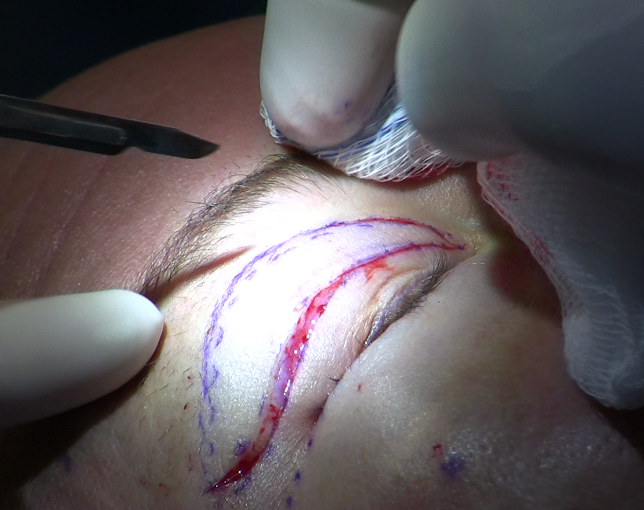
Upper blepharoplasty - surgical excision of excess skin, with management of the fat compartments as needed - is the only intervention that produces a lasting structural correction of dermatochalasis. Non-surgical options have a defined but narrow role.
Topical retinoids, radiofrequency skin tightening, and ablative laser resurfacing can improve skin texture and stimulate modest collagen remodeling in surface layers. They do not remove tissue, do not address herniated fat, and do not produce a clinically meaningful degree of correction in moderate or advanced dermatochalasis. They may serve as adjuncts for early-stage laxity or for maintaining skin quality after surgery, but they are not substitutes for tissue excision when true excess is present.
When concurrent true ptosis is found during the exam, the surgical plan needs to address both conditions. Ptosis repair changes the position of the lid margin, which in turn changes how the skin distributes across the lid - so the amount of skin to excise in a combined case differs from a pure dermatochalasis case. A surgeon who plans the combination correctly addresses ptosis first within the operative sequence, then determines skin excision based on the corrected lid height.
When the brow elevation test shows that significant brow ptosis is contributing to apparent lid excess, a brow lift may be indicated alongside or instead of blepharoplasty. Removing eyelid skin without addressing a clinically significant descending brow corrects the problem incompletely; the brow will continue pushing skin downward onto the lid after surgery.
What Surgery Cannot Fix if Only Skin Is Removed
The practical consequence of a missed pre-operative finding is not that the underlying problem goes permanently unaddressed. Most missed findings can be corrected in a second procedure. The constraint is that revision surgery after blepharoplasty operates with less available tissue than the primary operation did. Skin that has already been removed is gone, and the lid's reserve is reduced.
Some findings are recoverable with relatively straightforward revision. Herniated fat that was not managed at primary surgery can be approached through the original incision in a second procedure, though reopening a healed incision introduces scar tissue that changes the dissection planes. Concurrent ptosis that went unrecognized can be corrected by levator advancement without requiring additional skin excision, because ptosis repair does not depend on further reducing lid skin.
Missed brow ptosis is the harder situation. A brow that continues descending after blepharoplasty pushes skin onto a lid that now has a reduced reserve. Addressing the brow at that point is the correct next step, but attempting to remove more eyelid skin to chase the hooding the brow is actively creating risks reducing skin below the amount needed for comfortable lid closure - a functional problem that is difficult to reverse.
A separate consequence of incomplete pre-operative evaluation is asymmetry that surfaces after surgery. Two lids that appeared roughly symmetric before the procedure - because overhanging skin was obscuring differences in fat distribution or lid margin height - may reveal those differences once tissue is removed. That asymmetry was present anatomically before surgery; it was not created by the operation. But it becomes the presenting complaint at revision, and correcting it requires the same diagnostic precision the original evaluation should have applied.
Frequently Asked Questions
Does dermatochalasis get worse over time?
Yes. The collagen and elastic fiber loss driving the condition is cumulative and continues throughout life. Most patients notice a visible acceleration between their mid-40s and early 50s, when elastin synthesis drops and breakdown outpaces replacement. Surgery removes tissue that has already become excessive but does not stop the underlying biological process - the corrected lid continues to age on its new structural baseline. Patients with a strong genetic history or significant cumulative sun exposure tend to develop recurrent laxity more quickly than those whose condition appeared later and was primarily age-related. Most patients who have the procedure in their late 40s or 50s do not require revision within the first decade, though this varies by individual.
Can I tell from looking in a mirror whether I have dermatochalasis or ptosis?
You can get a rough sense. If eyelid skin folds over your lashes but both eyes appear open to roughly the same height, excess skin is the more likely finding. If one eye looks noticeably smaller or more closed, or if you find yourself habitually raising your brows to see better, true ptosis or a combined picture may be present. Only a clinical examination with MRD1 measurement and levator function testing can reliably separate them.
Is there anything that slows the progression before or after surgery?
The underlying process - collagen and elastic fiber degradation - is not reversible with any currently available topical or energy-based treatment. Broad sun protection slows UV-driven collagen breakdown and is a reasonable general measure. Active smoking is a documented independent risk factor and accelerates connective tissue aging throughout the body, including the eyelid. Beyond those modifiable factors, the rate of progression is largely determined by genetics. Topical retinoids may help maintain skin quality in early laxity but do not correct existing excess and have no effect on the fat compartments or the orbital septum. The same applies after surgery: the structural correction is lasting, but the underlying aging process continues on the corrected baseline.
What does the visual field test actually measure, and do I have to fail it to have surgery?
The visual field test maps where your vision is blocked by overhanging eyelid skin in the upper part of your visual field, with your lids in their natural resting position. Insurers who cover functional blepharoplasty require this documentation to establish that the dermatochalasis is obstructing vision, not only affecting appearance. If your results do not meet your insurer's current threshold, or if your condition is mild, surgery remains available as a cosmetic procedure - it is not technically different, but it is not covered by insurance.
Are there medical conditions that should be addressed before or alongside eyelid surgery?
Several systemic conditions are relevant to timing and surgical planning. Thyroid eye disease - most commonly associated with Graves disease - can produce orbital inflammation and tissue changes that continue after thyroid levels normalize; surgery performed during active disease or shortly after resolution may produce unpredictable results as orbital tissue continues to change. Significant pre-existing dry eye is also relevant, because blepharoplasty reduces available eyelid skin, and if the lid cannot fully close after surgery, dry eye symptoms can worsen. Anticoagulant use requires coordination with the prescribing physician before any elective procedure. These are standard items in a complete oculoplastic intake, and raising them at the initial consultation avoids delays in planning.
Are there conditions besides ptosis and brow ptosis that can look like dermatochalasis?
Several less common conditions can produce upper eyelid changes that resemble dermatochalasis or complicate its evaluation. Chronic contact dermatitis from cosmetics, contact lens solution, or topical eye medications can cause eyelid skin thickening and swelling that mimics early skin excess. Myasthenia gravis produces ptosis that fluctuates with fatigue and worsens over the course of the day, distinct from fixed skin excess but sometimes presenting alongside it. In younger patients, a palpable mass in the lid or an S-shaped lid contour may indicate a plexiform lesion requiring a separate evaluation before any blepharoplasty planning. Presentations with rapid onset, a palpable lid mass, marked asymmetry between the two eyes, or associated neurological symptoms warrant evaluation beyond the standard dermatochalasis workup before proceeding to surgery.
General information only, not medical advice. Individual anatomy and healing vary. See the disclaimer.