Every morning the eye is red, sticky, and full of grit. Drops help a little. By afternoon the irritation fades. The next morning it is back. For patients in this cycle, the standard workup - allergy testing, a new brand of artificial tears, removing contact lenses they may not even wear - produces nothing. The real culprit is structural: an upper eyelid that has lost the internal rigidity to hold its shape during sleep. It folds inside out against the pillow, grinds raw conjunctival tissue against fabric for hours, and flips back before the patient wakes. Floppy eyelid syndrome (FES) is the diagnosis those patients need, and understanding it properly means understanding a molecular process that connects a diseased eyelid to the sleep apnea the patient may not know they have.
The Tarsal Plate and What Happens When Elastin Disappears
The upper eyelid keeps its form because of the tarsal plate, a dense crescent of fibrous connective tissue that runs along the full width of the upper lid margin. It is roughly the width and curve of a thumbnail, and in a healthy eye it is stiff enough that everting the lid - flipping it inside out to expose the inner surface - requires deliberate lateral traction and both hands. The tarsal plate is what gives the lid its crisp, spring-loaded quality. Press it down and it returns immediately to the globe. Pull it away and it snaps back.
Threaded through that fibrous plate is a network of elastin fibers. Elastin is the structural protein responsible for elastic recoil - the property that allows tissue to deform under load and return to its original shape. When Culbertson and Ostler first formally described floppy eyelid syndrome in 1981, their case series documented something that had gone unrecognized: upper eyelids in obese men that could be everted effortlessly with minimal lateral traction and that spontaneously everted during sleep. The tarsal plate had lost its rigidity. Culbertson and Ostler hypothesized a loss of tarsal elasticity; subsequent histological studies confirmed what they had proposed, demonstrating that the elastin network inside the tarsus was markedly reduced or absent.
Without elastin, the plate becomes rubbery rather than firm. It deforms permanently under repeated mechanical stress and no longer springs back to its resting shape. This is not the same as skin becoming loose with age. The problem is deep inside the structural plate, not at the surface, and the cause is not gradual wear over decades but active enzymatic destruction driven by a systemic process.
MMP-7 and MMP-9: The Enzymes Dismantling the Tarsus
Immunohistochemistry of tarsal plate specimens from FES patients shows a consistent pattern: markedly reduced elastin density and elevated activity of matrix metalloproteinases MMP-7 and MMP-9. These enzymes are the specific molecular machinery responsible for degrading elastic tissue. Their presence at high concentrations in FES specimens is not incidental - they are the mechanism. They consume the elastin framework that keeps the tarsal plate stiff, and as that framework disappears, the plate loses all structural integrity.
Why Hypoxia-Reperfusion Drives the Same Cascade in Three Tissues
What triggers MMP upregulation? The answer connects the eyelid to the airway. During obstructive sleep apnea, oxygen saturation drops with each apneic episode and then rebounds as the airway reopens. That cycle of hypoxia followed by re-oxygenation generates oxidative stress - hypoxia-reperfusion injury - that activates a systemic inflammatory cascade and drives MMP production throughout the body. The same biochemical process that is consuming tarsal elastin is simultaneously degrading elastin in upper-airway soft tissue and elastic stroma in the cornea.
This is a systemic molecular disease with three visible endpoints in the same patient: loosening of the airway tissue that worsens OSA, dissolution of the tarsal plate that produces FES, and weakening of the corneal stroma that sets the stage for keratoconus. Understanding that the root cause is shared - not that one condition happens to coexist with another - is the conceptual shift that makes FES diagnostically and therapeutically coherent.
The MMP-driven molecular cascade that loosens upper-airway tissue in sleep apnea is simultaneously consuming the structural elastin of the eyelid and the cornea. Recognizing that shared biology is what transforms FES from a puzzling eye complaint into a diagnosable and treatable systemic process.
Who Develops Floppy Eyelid Syndrome
The original 1981 case series established a profile that still carries the strongest statistical weight: middle-aged obese men, many with undiagnosed obstructive sleep apnea. Male sex, middle age, and obesity are among the most consistently identified demographic risk factors across published series, and BMI above 30 is strongly associated in most studies. The mechanism is self-reinforcing: obesity promotes OSA, OSA drives MMP upregulation, and elevated MMP activity degrades both the tarsal plate and the airway soft tissue - which can worsen OSA further over time.
That profile is where to look first, but it is not where to stop. FES is increasingly recognized in women and in leaner, younger patients. The unifying feature across all of them is the hypoxia-reperfusion cycle, not body weight per se. A non-obese patient with severe positional OSA can develop FES by the same mechanism. The profile guides initial suspicion; it should not close the diagnosis to patients who fall outside it.
One demographic detail carries direct diagnostic weight: FES typically presents on the side the patient sleeps on. The lid that presses against the pillow absorbs the most direct mechanical stress - it everts against fabric during arousal events repeatedly across the night. A patient with right-sided symptoms who sleeps predominantly on their right side presents a pattern that bilateral dry eye disease or allergic conjunctivitis cannot explain. That single piece of history should redirect the workup.
Why FES Spends Years Disguised as Other Conditions
The symptoms patients bring to the office are nonspecific and morning-predominant, which means they match the appearance of several common diagnoses before anyone examines the lid mechanics:
- Redness that is worst on waking and clears gradually through the morning
- Mucous discharge or crusting along the lash line after sleep
- Gritty or foreign-body sensation that diminishes as the day progresses
- Papillary conjunctivitis of the superior palpebral conjunctiva with a cobblestone texture
- Chronic conjunctivitis that fails to respond to antibiotic or antihistamine therapy
- Photophobia and tearing that vary with which side the patient slept on
The slit-lamp hallmark - giant papillary conjunctivitis of the superior palpebral conjunctiva - is the finding that most reliably misdirects diagnosis. Large cobblestone papillae with whitish mucous discharge look exactly like contact-lens-related giant papillary conjunctivitis. The patient gets told to stop wearing lenses. If they do not wear lenses, they are told the papillae must be allergic. Neither treatment resolves the problem, because the cause is a mechanically damaged lid, not an immune reaction to contact-lens protein deposits or airborne allergens.
The temporal pattern is the most useful first filter. Irritation that is consistently worst immediately on waking and improves through the day is happening during sleep, not during waking exposure to environmental triggers. Dry eye tends to worsen as the day progresses. Allergy symptoms track with allergen exposure. Morning-predominant symptoms that ease without intervention point to a nocturnal mechanical source.
Making the Diagnosis: The Snap-Back Test and the Slit Lamp
There is no laboratory panel or imaging study that confirms FES. The diagnosis is made at the examination chair. Two findings together establish it: mechanical evidence of laxity and the characteristic conjunctival pathology.
The Snap-Back Test and Its Four Severity Grades
The snap-back test takes under a minute. The examiner draws the lid away from the globe and releases it, observing how the tissue returns. The response falls into one of four grades, and the grade directly determines whether conservative management or surgery is the appropriate next step.
| Grade | How the lid returns | Laxity level | Management direction |
|---|---|---|---|
| Normal | Immediate return to globe | None | No lid intervention needed |
| Mild | Slow return, without blink | Mild | Conservative measures first |
| Moderate | Returns only after a blink | Moderate | Conservative; surgery if it fails |
| Severe | Incomplete return even after blinking | Severe | Surgery strongly indicated |
After grading laxity, the examiner fully everts the upper lid under the slit lamp. Findings to look for include:
- Giant papillary conjunctivitis - cobblestone papillae that are often larger and more uniform than contact-lens-related papillae
- Whitish or mucoid discharge coating the papillae on the palpebral surface
- Superficial punctate keratopathy - small staining defects on the cornea from repeated nocturnal contact
- In advanced or long-standing disease: corneal vascularization, stromal haze, or frank scarring
- Microbial keratitis in the most severe cases where repeated nocturnal exposure has compromised the epithelial barrier
FES as a Gateway Diagnosis: Sleep Study and Corneal Screening
An FES diagnosis is not the endpoint of the workup. It is the starting point for two screenings that the patient may not have been offered before.
A systematic review and meta-analysis found obstructive sleep apnea in 57% of FES patients. For context, general adult OSA prevalence estimates vary considerably by diagnostic criteria and study population - older literature using a symptomatic definition commonly cited figures around 2 to 7%, while broader current estimates using an apnea-hypopnea index threshold of 5 or more run as high as 10 to 30% depending on the population studied. By any of these benchmarks, the prevalence of OSA in FES patients is substantially and consistently elevated. Every patient diagnosed with FES should be referred for polysomnography or a home sleep apnea test regardless of whether they report snoring, witnessed apnea, or daytime sleepiness. Many OSA patients have no idea they stop breathing during the night.
The corneal risk is less widely known but carries serious implications. A case-control study found an odds ratio of 19.3 for keratoconus in FES patients compared with age-matched controls. That is not a modest statistical association - it is a near-20-fold increase in risk. The mechanism is the same MMP-7 and MMP-9 activity that stripped elastin from the tarsus; corneal stroma is similarly elastin-rich, and the same enzymatic process weakens its architecture. A patient with FES who develops progressive keratoconus without surveillance may lose substantial vision before the corneal condition is caught at an actionable stage.
At the same visit, the following referrals and tests should be initiated:
- Sleep medicine referral for polysomnography or home sleep apnea test
- Corneal topography to establish a baseline and screen for early keratoconus
- Cardiovascular risk assessment - untreated OSA carries independent cardiovascular burden
Conservative Treatment: The Sequence Before Surgery
Mild-to-moderate laxity and all patients waiting for OSA workup results should begin conservative management immediately. The goal is to remove the nocturnal mechanical insult, reduce systemic MMP activity, and protect the ocular surface while severity is being established.
- Treat confirmed OSA with CPAP: Reducing nightly hypoxia-reperfusion cycles lowers systemic MMP activity and decreases the arousal-related mechanical rubbing that everts the lid. Published studies consistently document meaningful improvement in ocular surface health in a substantial portion of FES patients once OSA is treated, though reported improvement rates vary across series - check current literature for the most recent figures. Start here when sleep study results are available.
- Adjust CPAP mask fit for air leaks: If the patient reports eye irritation attributable to mask air escaping toward the cornea, have the sleep equipment provider refit the mask or switch to a different mask style. This is a common problem with a practical fix that does not require abandoning CPAP.
- Apply lubricating ointment at bedtime: A thick ointment formulation - not a thin drop - creates a surface barrier between the exposed conjunctiva and the pillow in the event the lid everts during sleep.
- Tape the lateral lid or use an eye shield at night: A firm ocular shield taped over the closed eye, or a strip of tape drawn lightly across the lateral lid margin before sleep, physically prevents eversion. This typically produces the most immediate symptom relief of any conservative measure.
- Reposition with a cylindrical pillow: A roll or cylindrical pillow supports the cheek so the orbit clears the mattress surface, reducing direct contact between the lid margin and bedding. It does not correct laxity but removes the primary mechanical insult during sleep.
Success with conservative management means morning symptoms resolve substantially and snap-back grade remains stable or improves. Patients whose symptoms persist after six months of consistent conservative management, and those whose corneal findings are progressing, should be offered surgery rather than accumulating further corneal damage.
Lateral Tarsal Strip Surgery: What It Actually Does
The lateral canthal tendon is the fibrous attachment that anchors the outer corner of the eyelid to the lateral orbital rim - the bony wall at the outer edge of the eye socket. In FES, this tendon has lost tensile integrity along with the tarsal plate. The lid hangs loosely at its outer corner. Lateral tarsal strip surgery corrects this by constructing a new, firm anchor between the tarsal plate and the bone.
The Anatomy of the Repair
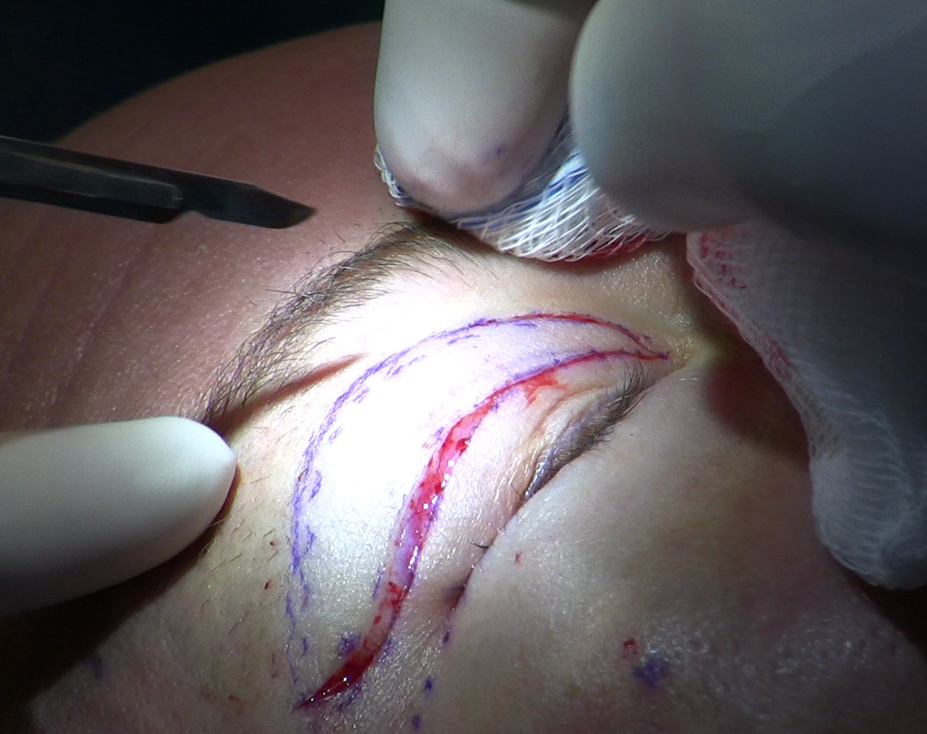
The procedure is performed under local anesthesia with sedation in an outpatient setting. The surgeon separates the upper and lower limbs of the lateral canthal tendon at the outer canthus, exposing the end of the tarsal plate in the upper lid. The overlying conjunctiva and anterior skin-muscle layer are removed from the terminal portion of the plate, leaving a clean strip of bare tarsus - the lateral tarsal strip that gives the procedure its name. This strip is then passed through the wound and sutured directly to the inner surface of the periosteum on the lateral orbital rim, at the anatomically correct height to reproduce the natural canthal angle. Excess tarsal tissue is trimmed, and the outer canthus is reconstructed.
The reliability of the repair comes from the quality of what is being joined: firm tarsal plate tissue is anchored to rigid bone at a defined anatomical landmark. There is no reliance on stretched tendon or on skin, both of which can re-elongate. Published series report 91 to 98% long-term symptom resolution, and that range is consistent across series because the procedure addresses the root mechanical failure.
When laxity involves both the outer and inner corners of the lid - which occurs in more severe and long-standing cases - medial canthopexy is added to reinforce the medial canthal attachment as well. In a published series of 18 consecutive patients treated with combined medial canthopexy and lateral tarsal strip, all 18 achieved symptom relief and objective resolution of lid laxity. Not every FES patient needs medial canthopexy; the decision depends on snap-back grade, symptoms, and intraoperative assessment of medial tension.
Long-Term Monitoring After an FES Diagnosis
Surgical lid tightening corrects the mechanical problem it targets. It does not reverse the underlying MMP biology, does not cure OSA, and does not eliminate corneal risk. Long-term management is ongoing and involves more than the eyelid.
Corneal topography should be obtained at baseline and repeated at least annually, with more frequent examinations if any topographic irregularity is present. The goal is to catch keratoconus while corneal cross-linking - a procedure that stiffens the cornea and halts progression - remains a viable option. Patients referred for corneal transplant because their keratoconus was discovered late represent a failure of the monitoring plan, not an inevitable outcome.
The full long-term surveillance framework includes:
- Annual corneal topography, with baseline obtained at the time of FES diagnosis
- Ongoing OSA management review - CPAP adherence directly affects the rate of continued MMP-driven tissue degradation
- Lid laxity reassessment if symptoms recur - tarsal strip surgery is durable, but patients with active, poorly controlled OSA and continued mechanical stress can develop recurrent laxity over years
- Ocular surface evaluation at each visit for punctate keratopathy or new corneal vascularization, which signal inadequate protection
FES is a condition that connects oculoplastic surgery, sleep medicine, and corneal specialty care. A patient managed in only one of those tracks has an incomplete care plan. The oculoplastic surgeon addresses the lid. The sleep medicine specialist controls the driving biochemical process. The corneal specialist watches the tissue that is most vulnerable to the downstream consequences. All three roles matter.
Frequently Asked Questions
Can floppy eyelid syndrome affect both eyes at the same time?
Yes, bilateral FES occurs and is more common in patients with severe, untreated OSA and high BMI, where systemic MMP activity affects both tarsal plates equally. Unilateral presentation is more typical and nearly always corresponds to the patient's preferred sleeping side. Symmetric bilateral disease can be harder to distinguish from dry eye on history alone, which makes the snap-back test the critical distinguishing step.
Is floppy eyelid syndrome the same as ptosis?
No. Ptosis is drooping caused by weakness or detachment of the levator muscle or its aponeurosis - the lid sits too low because the elevator mechanism is impaired. FES involves a lid that may sit at normal height when the patient is awake and upright, but that has lost internal structural rigidity and everts effortlessly when tested or when the patient lies down. Levator function is typically preserved in FES. The two conditions can coexist, but they are distinct diagnoses with different treatments.
Will treating sleep apnea make surgery unnecessary?
For patients with mild snap-back grades, CPAP therapy combined with conservative measures may resolve morning symptoms sufficiently without an operation - published series consistently show that a meaningful portion of FES patients see ocular surface improvement with OSA treatment, though improvement rates vary by series. Severe laxity rarely reverses with CPAP alone because the tarsal plate has already lost structural integrity; the biochemical process can be slowed, but the plate will not rebuild elastin on its own. OSA treatment and surgery are most often complementary interventions rather than competing choices.
I have giant papillary conjunctivitis but do not wear contact lenses. Could this be floppy eyelid syndrome?
That is exactly the presentation pattern that should prompt an FES evaluation. Giant papillary conjunctivitis without a contact lens, ocular prosthesis, or exposed suture as the mechanical driver is a strong pointer toward FES. Ask your ophthalmologist to fully evert the upper lid, examine it under the slit lamp, and perform a snap-back test. Diagnosing this correctly stops years of treatment for the wrong condition.
My corneal topography shows early irregular astigmatism - could FES be part of the explanation?
It could, particularly if you also have morning-predominant eye irritation, sleep on the side of the affected eye, or have known or suspected OSA. The roughly 19-fold elevated odds of keratoconus in FES patients reflects a real shared mechanism - the same MMP enzymes that degrade tarsal elastin degrade corneal stroma. If FES has not been evaluated, a lid laxity assessment alongside your corneal monitoring adds important diagnostic information and may identify a controllable driver of corneal progression.
How long does recovery take after lateral tarsal strip surgery?
Bruising and swelling at the outer corner of the eye are normal for one to two weeks. The reconstructed canthus fully heals and the lid position stabilizes over approximately four to six weeks as the sutured tarsal strip integrates with the periosteum of the orbital rim. Many patients notice that morning irritation improves within the first days after surgery because the mechanical source of nocturnal exposure is immediately addressed. Final assessment of the outcome is typically done at the three-month mark.
General information only, not medical advice. Individual anatomy and healing vary. See the disclaimer.