Chronically watery eyes send most patients down a long road of antihistamines and artificial tears before anyone looks at where the tears actually go. The drainage system that pulls tears from the eye into the nose has four distinct anatomical levels, and a problem at any one of them produces the same symptom - constant overflow - but demands a completely different fix. More surprising still: in roughly four out of ten patients who see an oculoplastic specialist for this complaint, the problem is not a blockage at all. Understanding the anatomy, the causes at each level, and the diagnostic steps that separate them is the fastest route from a permanently wet cheek to a dry one.
Why Eyes Make Tears and How They Drain: The Four-Level Anatomy Every Patient Should Know
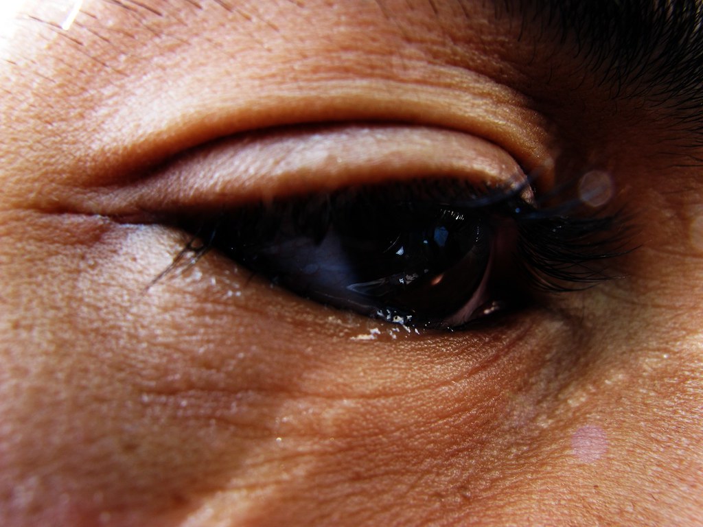
Tears are produced continuously by the lacrimal gland above the upper eyelid and by small accessory glands across the conjunctiva. Each blink spreads the film across the eye and pushes tears toward the medial corner - the inner corner near the nose - where they enter the drainage system. Four sequential structures carry them from there into the nasal cavity.
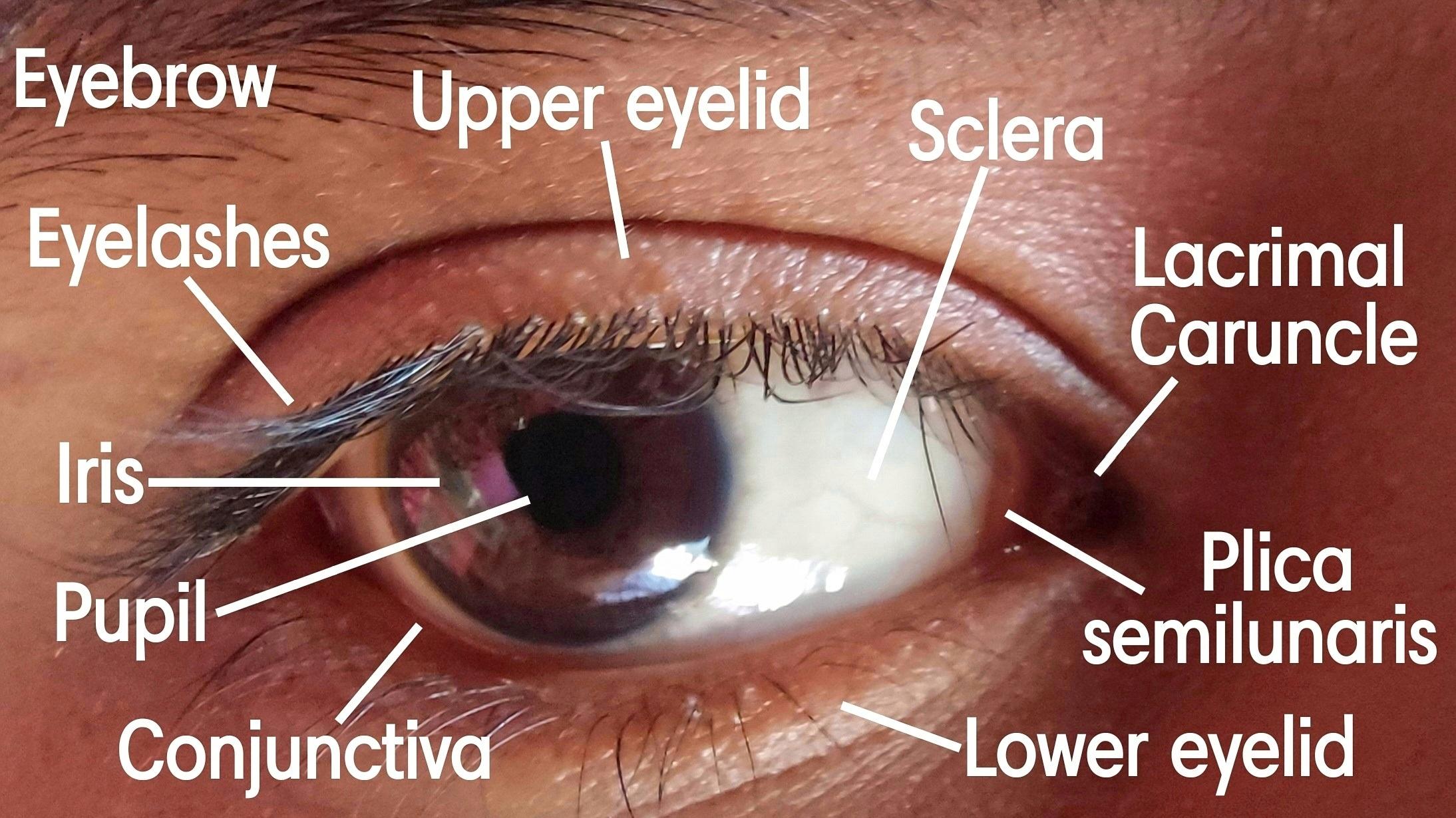
- Punctum: A small round opening on the margin of each eyelid at the inner corner - one upper, one lower. Tears enter here first. The punctum must be open, correctly positioned, and in contact with the tear lake for drainage to begin.
- Canaliculi: Narrow tubes that run along each lid margin from the punctum toward the nose. The upper and lower canaliculi travel separately for roughly 8 mm, then merge into a common canaliculus. At this junction sits the valve of Rosenmüller, a mucosal fold that acts as a one-way check to prevent backward reflux.
- Lacrimal sac: A small reservoir seated in a bony depression - the lacrimal fossa - along the medial orbital wall. The sac collects drainage from the canaliculi and passes it downward.
- Nasolacrimal duct: A bony channel that carries tears from the lacrimal sac into the nasal cavity, emptying under the inferior turbinate. This is why crying makes the nose run.
The system does not run on gravity alone. The orbicularis oculi muscle, encircling the eye, creates an active pumping action with every blink - compressing the canaliculi and sac to draw tears inward. When that muscle is weak, or when the lower eyelid is loose, the pump fails even if every structure from punctum to nasolacrimal duct is anatomically open.
The Dry-Eye Paradox: When Watery Eyes Are Caused by Too Little Tear Film
The most counterintuitive finding in the oculoplastic literature on watery eyes is this: approximately 40% of patients presenting to a specialist with epiphora have dry eye as the underlying driver, not a blocked drain. When the baseline tear film is inadequate - from meibomian gland dysfunction, aqueous deficiency, or goblet cell loss - the corneal surface becomes irritated, and the nervous system responds by triggering the lacrimal gland to flood the eye with reflex tears.
Reflex tears differ from baseline tears. They are predominantly watery, lacking the lipid and mucin layers that stabilize the film. They overwhelm the drainage system, spill onto the cheek, then evaporate quickly - leaving the cornea irritated again. The cycle restarts. A patient with severe dry eye can spend the day wiping a wet face while their eye is simultaneously starved of a stable film.
This distinction matters enormously for treatment. A patient in this group does not need lacrimal surgery - they need dry-eye treatment. Aggressive lubrication, omega-3 supplementation, and management of eyelid margin disease often eliminate the tearing entirely without any drainage procedure.
Eyelid Position Problems: How Ectropion, Laxity, and Pump Failure Cause Tearing Without Any Blockage
Even a fully patent, unobstructed drainage system cannot work if the eyelid mechanics that feed it are broken. Three distinct problems fall into this category, and each has a different solution.
- Ectropion: Outward rolling of the lower eyelid displaces the punctum away from the tear lake. Tears pool at the lid margin but cannot enter because the punctum no longer contacts the fluid. The fix is surgical repositioning of the lower lid itself - not a lacrimal procedure. Once the lid is back against the globe, the punctum returns to the tear lake and drainage resumes normally.
- Lower eyelid laxity: The lid may not be overtly everted, but age-related looseness allows it to sag slightly at the medial portion. The punctum points inward but no longer makes tight contact with the eye, and the capillary action that draws tears into the opening is lost.
- Lacrimal pump failure: In patients with facial nerve palsy, the orbicularis muscle is denervated and the blink is incomplete or absent - tears spill because the pump never fires. The same mechanism operates in floppy eyelid syndrome, where chronic horizontal lid laxity undermines the mechanical efficiency of every blink. These patients need treatment aimed at restoring eyelid function, not at the drainage apparatus.
Punctum Problems: Stenosis, Malposition, and Which Medications Silently Narrow the Drain
The punctum is the entry point of the drainage system and the level most vulnerable to medication side effects. Several drug classes cause clinically significant punctal narrowing that goes unrecognized for years.
- Topical anti-glaucoma drops - particularly prostaglandin analogues - cause goblet cell loss on the conjunctival surface and progressive fibrosis of the punctal rim with long-term use.
- Systemic chemotherapy with taxanes, especially docetaxel, causes punctal and canalicular stenosis. Timing matters: weekly administration schedules cause this significantly more often than every-three-week schedules. Patients on weekly taxane regimens should be screened for early lacrimal narrowing before symptoms become severe.
- 5-fluorouracil independently causes punctal and canalicular fibrosis through similar mechanisms.
- Chronic blepharitis - long-standing inflammation at the eyelid margin - produces gradual scarring at the punctal opening over years.
Punctal stenosis from any cause mimics a blocked nasolacrimal duct symptomatically. The level of obstruction is different and the treatment is much simpler - a small procedure at the punctum itself, not a major operation.
Canalicular Obstruction: The Underdiagnosed Infection, Trauma Repairs, and Drug-Induced Scarring
Canaliculitis is one of the most consistently misdiagnosed conditions in oculoplastic practice. The most commonly cited causative organism is Actinomyces israelii, a filamentous gram-positive bacterium that colonizes the canalicular tube and forms hard yellow concretions - sulfur granules - inside it; other bacteria and polymicrobial infections also occur and should be considered in culture-positive cases. Patients typically present after months or years of unilateral discharge and tearing, having already received multiple courses of antibiotic eyedrops for presumed conjunctivitis, with little effect.
The clinical clues are specific: the affected punctum appears pouting and slightly swollen, there is yellow or gritty discharge expressible from it, and the canaliculus feels thickened and firm on palpation. Antibiotic drops cannot eradicate concretions - they treat the surface without reaching the intracanalicular source. Definitive treatment is canaliculotomy: a small incision to open the canaliculus, followed by curettage to remove the material. Patients who have this done after years of misdiagnosis frequently find it immediately resolves the problem that nothing else touched.
Canalicular laceration from facial trauma - a dog bite, a hook, a blow to the medial face - requires surgical repair over a silicone stent to prevent permanent obstruction. The window is short: repair within 24-48 hours of injury gives the best outcomes. After five to seven days, inflammatory scarring begins, re-anastomosis becomes technically much harder, and the risk of lifelong epiphora on that side rises substantially.
Lacrimal Sac and Nasolacrimal Duct Obstruction: The Most Common Acquired Cause and Who Gets It
Obstruction at the level of the lacrimal sac or nasolacrimal duct is the most common cause of acquired epiphora requiring surgery in adults. It develops through gradual fibrosis - scar tissue progressively narrows the lumen until flow stops. Fibrosis can involve the junction of the common canaliculus with the lacrimal sac, the lacrimal sac wall itself, or the nasolacrimal duct; in many patients more than one level is affected. Once the nasolacrimal duct itself is involved, dilation cannot restore flow - a new drainage route must be created.
Women develop acquired nasolacrimal duct obstruction more often than men. The reason appears structural: females have anatomically narrower nasolacrimal ducts, leaving less margin before age-related or inflammatory fibrosis narrows the channel to failure. The condition is most common after age 40 and increases in frequency with age.
Congenital Obstruction in Infants
A separate population with nasolacrimal duct obstruction is newborns, where the duct fails to fully open at birth. The natural history is encouraging: the majority of these cases resolve spontaneously or with massage by 12 months of age, with published resolution rates exceeding 90% in most series. Probing success rates are generally favorable when performed in the first year of life, though published figures vary widely across studies and by technique - rates of roughly 85-98% have been reported before 12 months depending on study design and setting. Success rates decline with increasing age, and published figures for children over 24 months range from roughly 70-90% across individual studies; probing in older children is typically performed under general anesthesia. Delays trade a simple probe for a more involved procedure. Children who fail probing beyond 18 months are candidates for silicone tube intubation or, in persistent cases, DCR.
How the Diagnosis Is Made: Dye Disappearance Test, Irrigation, and When Imaging Adds Useful Information
Diagnosing epiphora is a stepwise process that works from the outside in. The history - duration, which eye, current medications, prior chemotherapy, any facial trauma - directs the examination before any instruments are used.
- Assess eyelid position and tone. Is there ectropion? Is the lower lid lax? Does the patient have facial palsy or difficulty closing the eye? These findings direct treatment before any dye is used.
- Examine the punctum directly. Is it visible, open, and contacting the eye? Does it appear inflamed or pouting? Can discharge be expressed with pressure over the canaliculus?
- Perform the fluorescein dye disappearance test. A drop of fluorescein is instilled and the eye is examined under cobalt-blue light at five minutes. Complete clearance argues against significant drainage obstruction - the tearing is likely from overproduction or a pump problem. Persistent dye confirms drainage failure.
- If the dye test suggests obstruction, lacrimal irrigation follows. A blunt cannula is introduced through the punctum and saline is irrigated. Whether fluid passes into the nose, refluxes back through the same punctum, or returns through the opposite punctum localizes the obstruction to a specific anatomical level.
- Imaging is reserved for cases where a lacrimal sac mass is suspected, for revision surgery planning, or when syringing results are equivocal. CT dacryocystography outlines the anatomy and can identify sac distension, filling defects, or bony abnormalities.
Non-Surgical and Minor Surgical Fixes: Dilation, Punctoplasty, and Intubation
Not every patient needs a major operation. When obstruction is at the punctum or involves mild canalicular narrowing, simpler procedures achieve lasting results.
Punctal dilation uses a tapered metal dilator introduced through the punctum to widen a narrowed opening. It is a two-minute procedure requiring only a topical anesthetic drop. For mild stenosis it gives immediate relief, but fibrosis tends to recur without further treatment.
Punctoplasty is more durable. The surgeon creates a small incision to permanently enlarge the punctal opening, removing the fibrotic tissue causing narrowing. When combined with a temporary silicone stent that holds the opening patent during healing, published studies show functional resolution of tearing in roughly 79-84% of patients. Controlled comparisons show simple dilation performs comparably to the Kelly punch punctoplasty technique, suggesting the stent - not the specific cutting method - drives the result.
Silicone intubation alone is used for mild canalicular obstruction or as an adjunct to scaffold a newly opened segment during healing. Stents are left in place for three to six months, then removed in a brief office visit with no anesthesia needed.
Dacryocystorhinostomy (DCR): What the Operation Actually Does, External vs. Endonasal, and What Recovery Looks Like
When obstruction involves the lacrimal sac or nasolacrimal duct, dilation and stenting cannot restore drainage - the blocked segment cannot be stretched open. Dacryocystorhinostomy bypasses the problem entirely by creating a small window directly between the lacrimal sac and the nasal cavity, giving tears a new route that sidesteps the obstructed duct permanently.
| Criteria | External DCR | Endonasal DCR |
|---|---|---|
| Approach | Small skin incision at medial canthal area | Entirely through nasal cavity, no skin incision |
| External scar | Approximately 10 mm near the nose bridge | None |
| Operative time | Longer in most comparative studies | Shorter in most comparative studies; reported times vary widely by series and surgeon |
| Intraoperative bleeding | Higher; comparative studies consistently show more intraoperative bleeding with external DCR than endonasal, though published rates vary widely between series | Lower; comparative studies consistently show less intraoperative bleeding with endonasal DCR than external, though published rates vary widely between series |
| Published success | High - 85-99% in the literature | High - 80-96% in the literature |
| Preferred when | Lacrimal sac tumor suspected; revision after failed endonasal; prior medial canthal scarring | Concurrent nasal pathology; patient preference for no scar; prior medial canthal surgery limiting external access |
A UK study of more than 500 consecutive operations found equivalent outcomes at one year for both approaches in primary nasolacrimal duct obstruction, making the choice largely patient- and surgeon-specific rather than outcome-driven.
The choice between external and endonasal DCR should be driven by the underlying pathology, concurrent nasal disease, and the surgeon's experience - not by an assumption that one approach is universally superior. Outcomes at one year are comparable in head-to-head published data.Synthesis of comparative DCR outcome literature
Recovery and What to Expect
DCR is typically performed under general anesthesia or intravenous sedation and completed as a day procedure. Nasal congestion and mild bloody nasal discharge are common in the first week. External DCR produces bruising and swelling around the medial eye that generally resolves within two weeks. Patients are advised to avoid nose-blowing for the first week to prevent air from tracking through the new ostium and causing lid swelling. The eye continues to water during healing - this is not a sign of failure. Most patients notice clear improvement within four to six weeks; final assessment follows stent removal, which is done in the office without anesthesia.
Frequently Asked Questions
How do I know whether my watery eye is dry eye or a blocked duct?
The fluorescein dye disappearance test distinguishes the two in the office: complete clearance within five minutes argues against significant drainage obstruction, pointing toward overproduction. Dry eye typically also produces burning, grittiness, and symptoms that worsen in wind or air conditioning. An oculoplastic evaluation will assess both possibilities before any treatment is planned.
Can a blocked tear duct clear on its own in adults?
Spontaneous resolution is common in infants but uncommon in adults with acquired nasolacrimal duct obstruction. In adults, fibrosis at the duct level is progressive - the obstruction typically worsens without treatment. Mild narrowing caught early may respond to dilation, but a complete obstruction in an adult generally requires DCR for lasting relief.
Is DCR permanent, or will the blockage come back?
DCR creates a new bony opening between the lacrimal sac and the nasal cavity - the original blocked duct is bypassed, not reopened. The new ostium can close through fibrosis, particularly if the silicone stent is removed early or if significant nasal mucosal disease is present. When done correctly with appropriate stent placement, published success rates are high, with the majority of patients maintaining free drainage long-term. A small percentage require revision.
My eye has been watering for years but only on one side - what does that mean?
Unilateral epiphora is clinically significant. It points to a structural or infectious cause on that specific side rather than a systemic condition like dry eye, which usually affects both eyes. Unilateral chronic tearing - especially with any medial canthal swelling or discharge - warrants evaluation to rule out lacrimal sac obstruction, canaliculitis, or, less commonly, a lacrimal sac mass requiring imaging before any procedure.
I take glaucoma drops - could they be causing my watery eyes?
Possibly, and through two separate mechanisms. Topical glaucoma medications can cause punctal fibrosis and ocular surface irritation that triggers reflex tearing; they can also impair the goblet cells that stabilize the tear film, initiating the dry-eye cycle. If you use topical glaucoma drops and have epiphora, tell your oculoplastic surgeon - the drops will factor into the workup and may need to be managed in coordination with your glaucoma specialist before any lacrimal procedure is planned.
What happens if a canalicular tear from an injury is not repaired quickly?
The repair window is narrow. Within 24-48 hours, re-anastomosis over a silicone stent is straightforward and carries a high chance of restoring full drainage. After five to seven days, the cut ends begin to contract and scar, making repair technically harder and raising the risk of permanent epiphora on that side. Any laceration near the inner corner of the eyelid - from a dog bite, a hook, or blunt facial trauma - should be evaluated by an oculoplastic surgeon the same day or the following morning, not at a routine follow-up appointment.
General information only, not medical advice. Individual anatomy and healing vary. See the disclaimer.