Before a surgeon recommends any approach to lower eyelid surgery, one anatomical question takes priority over technique, incision location, or patient age: where does the cheekbone sit relative to the eye? That relationship - the orbital vector - determines whether the lower eyelid has solid bony support beneath it or is essentially cantilevered over empty space. In patients where the eye protrudes past the cheekbone, the standard lower blepharoplasty plan carries risks that do not exist in patients with different anatomy. Those risks are not eliminated by choosing a gentler technique. Understanding what orbital vector means, how surgeons find it in the clinic, and what it requires of the surgical plan is the foundation for any informed conversation about lower eyelid surgery.
What Orbital Vector Means
The lower eyelid maintains its position through a combination of structures: the canthal tendons anchoring it laterally and medially, the orbicularis muscle, the tarsal plate, and - less obviously - the passive support of the bony ledge below. The cheekbone, specifically the malar eminence and the infraorbital rim, normally projects forward far enough that the lower lid has something to rest against. When that bony shelf is set far enough forward, gravity and the weight of soft tissue work with the system rather than against it.
The orbital vector describes the spatial relationship between the anteriormost point of the globe - the cornea - and the anterior face of the cheekbone, assessed from the side. The check is straightforward: standing in profile, a surgeon drops an imaginary vertical line from the cornea straight downward and notes where it lands relative to the malar eminence. That single observation tells a great deal about what the lower eyelid is working with structurally.
Positive, Neutral, and Negative: What Each Means for the Lower Lid
The three orbital vector types produce different mechanical conditions for the lower eyelid, and they carry different implications for surgical planning.
Positive Vector
The malar eminence sits ahead of the cornea in the profile view. The bony ledge provides clear anterior support. The lower lid has something to rest against, and mild amounts of surgical manipulation - fat removal, skin excision, incision placement - are tolerated well because the underlying structure is working in the lid's favor. Most patients with a clearly positive vector handle lower blepharoplasty across a range of techniques without elevated lid malposition risk.
Neutral Vector
The cornea and cheekbone are roughly coplanar. Support is present but less generous. These patients are not high risk by default, but a surgeon will independently evaluate lid laxity and consider canthal reinforcement if any laxity is found on examination.
Negative Vector
The cornea projects forward past the cheekbone. The lid has no solid bony support anterior to the globe. It relies entirely on soft tissue attachments to maintain contact with the eye. A negative orbital vector is the most consistently identified preoperative anatomical risk factor for lower eyelid retraction after blepharoplasty. The following features tend to accompany it:
- Prominent or forward-sitting eyes relative to the cheek profile
- A deep tear trough or hollow under-eye appearance
- Visible sclera (white of the eye) below the iris even before any surgery
- A flattened or recessed cheek below the eye when viewed from the front
- History of needing to blink to keep the lower lid fully against the eye
What Creates a Negative Orbital Vector
Four distinct underlying causes produce this anatomy, and they differ in mechanism, in what the skeleton contributes, and in what surgical options realistically exist.
| Cause | Mechanism | Skeleton involved | Key surgical implication |
|---|---|---|---|
| Skeletal midface hypoplasia | Infraorbital rim and anterior maxilla underdeveloped | Yes - primary deficit | Rim augmentation can directly correct the deficiency |
| High axial myopia | Elongated globe protrudes relative to a normal cheekbone | No - skeleton is typical | No skeletal fix available; all modifications are soft-tissue and canthal |
| Thyroid eye disease | Orbital inflammation pushes globe forward (true proptosis) | No - skeleton is typical | Disease must be fully stable before any elective lid surgery |
| Age-related maxillary recession | Anterior maxilla involutes over decades; rim migrates posteriorly | Yes - acquired deficit | Volume loss and lid laxity often coexist; combined approach usually required |
Skeletal midface hypoplasia means the infraorbital rim and anterior maxilla simply did not develop fully. The bony support structure was set back from early life. High axial myopia is a different problem: the skeleton can be completely normal, but an axial eye length past the threshold that defines significant myopia means the elongated globe protrudes relative to the cheekbone. Published case reports have documented this in patients who required modified lower blepharoplasty because of myopia alone - including in identical twins, where the shared anatomy confirmed the mechanism clearly.
Thyroid eye disease produces true proptosis through orbital inflammation that physically pushes the globe forward. Because the vector disturbance is driven by active disease rather than fixed anatomy, operating while inflammation is present or recently resolved risks adding a surgical insult to an unstable situation. The timeline for surgical candidacy in this group requires close coordination with the treating ophthalmologist or endocrinologist.
Age-related maxillary recession is the cause most often missed because it develops slowly and without any single identifiable event. The facial skeleton is not static across a lifetime. As the anterior maxilla involutes, the infraorbital rim migrates posteriorly while the globe holds its position. A person with a neutral vector at forty may have a clearly negative one by sixty-five - not because the eye moved forward, but because the bone beneath it gradually retreated. This is a common contributor in older patients seeking lower eyelid rejuvenation who have no prior surgical history and no eye disease.
How a Surgeon Assesses Vector in the Office
The orbital vector check requires no instruments. A surgeon observing the patient's profile drops an imaginary vertical line from the cornea: if the cheekbone lies at or ahead of that line, the vector is positive or neutral; if the cheekbone falls behind it while the cornea projects forward, the vector is negative. This visual assessment takes seconds and should occur at every preoperative lower blepharoplasty consultation without exception.
Two physical tests then quantify the mechanical state of the lower eyelid itself - its laxity - which becomes critical information once a negative vector is confirmed.
The Snap Test
The surgeon pulls the central lower lid directly away from the globe and then releases it without prompting the patient to blink. A normal lid snaps back into contact with the globe in under one second. An abnormal result is the lid taking longer than one second to return, or requiring a blink to complete the return. A slow snap indicates lid laxity - the soft tissue attachments are already stretched or weakened - which compounds the retraction risk created by the vector anatomy.
The Distraction Test
The surgeon pulls the lower lid directly away from the cornea horizontally and measures the travel distance. Less than 2 mm is normal. More than 6 mm indicates significant canthal tendon laxity. More than 8 mm is clearly abnormal and signals that horizontal support is substantially compromised. In a negative vector patient, even moderate distraction findings push the surgical plan toward canthal reconstruction rather than simple reinforcement. Together, these three assessments form a complete preoperative orbital evaluation using only clinical observation and manual examination.
What the Research Shows
The connection between negative orbital vector and lower eyelid retraction is documented, not inferred. A cross-sectional study of 123 individuals aged 20 to 80 divided subjects into those with normal lower eyelid position and those showing lower eyelid retraction - with no prior surgery in either group. The subjects with normal lids had a mean orbital vector angle of +9.76 degrees. The subjects who already showed retraction had a mean of -13.65 degrees.
Negative orbital vector anatomy predisposes the lower eyelid to retraction before any surgical instrument is involved - the malposition risk is structural, present in unoperated eyes, and built into the anatomy rather than created by an operation.Cross-sectional study of 123 subjects, PMC7276168
This finding matters because it reframes the problem entirely. Negative orbital vector is not primarily a surgical complication - it is a preexisting anatomical condition that surgery can worsen if not accounted for. If the geometry alone produces lid retraction in eyes that have never been touched, then operating on those same eyes without modifying the plan adds a further destabilizing force to a system already at its limit.

Why Standard Lower Blepharoplasty Carries Specific Risks in Negative Vector Patients
Lower eyelid malposition - scleral show, retraction, or ectropion - is the single most common complication after lower blepharoplasty. In negative vector patients, the anatomical conditions for that complication exist before the first incision. Standard approaches that work well in positive or neutral vector patients apply forces to a lower lid that was already compensating.
The risks increase further when skin excision enters the plan. Removing lower eyelid skin shortens the anterior lamella - the skin is now tighter than before. In a well-supported eye, moderate skin excision is typically tolerated. In a negative vector patient, the same degree of shortening pulls an already-vulnerable lid downward and outward. The resulting retraction or ectropion causes chronic dryness, exposure-related corneal irritation, and an asymmetric appearance that is both medically and aesthetically significant. Correcting it requires revision surgery - often involving spacer grafts, canthal reconstruction, and midface lifting - that is far more involved than the original operation.
Surgical Modifications That Reduce Risk
Treating a negative vector patient does not mean performing standard blepharoplasty more carefully. The plan itself changes in several ways, each addressing a specific mechanical weakness.
- Canthal support is mandatory. Canthopexy - tightening and repositioning the existing lateral canthal tendon - or canthoplasty - reconstructing the tendon through a new fixation point on the orbital rim - must be included in every negative vector lower blepharoplasty. Performing the operation without canthal support in this anatomy substantially raises retraction risk. Canthopexy is appropriate when existing tendon tissue is adequate; canthoplasty is required when laxity findings on the distraction test indicate the tendon itself is insufficient.
- Fat repositioning over fat excision. Rather than removing herniated orbital fat, the surgeon repositions it - advancing it over the infraorbital rim to fill the tear trough while keeping the volume in place behind the lid. This preserves the passive upward pressure fat exerts on the lid from behind, avoids the hollow appearance that makes globe prominence worse after excision, and adds soft-tissue volume along the rim where it improves both support and contour.
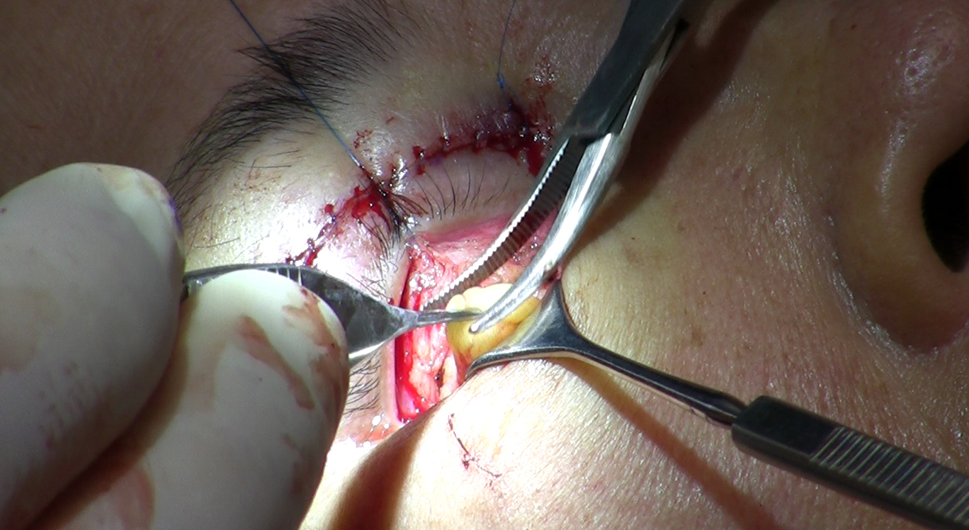
- Transconjunctival approach combined with a SOOF lift. The sub-orbicularis oculi fat lift elevates the midface fat compartment upward, simultaneously filling the tear trough and adding vertical soft-tissue support to the lower lid from below. Combined with the transconjunctival incision - which avoids disrupting the anterior lamella entirely - this approach addresses the support deficit from a direction that standard blepharoplasty does not reach.
- Minimal or deferred skin excision. Any reduction in lower eyelid skin length acts against a lid that is already working to maintain globe contact. In moderate to severe negative vector cases, skin removal may be deferred until the structural situation is addressed or avoided entirely in favor of resurfacing techniques that tighten without shortening the lamella.
Addressing the Skeleton: When the Bone Position Is the Real Problem
Soft-tissue surgery cannot move bone. Any combination of fat repositioning, canthal tightening, and SOOF lifting leaves the infraorbital rim in the same position it occupied before the operation. In patients whose negative vector comes primarily from skeletal underdevelopment or age-related maxillary recession - rather than from globe protrusion - the structural deficit is in the bone, and soft-tissue work operates around a problem it cannot solve.
Injectable Fillers as a Test and as a Treatment
Filler placed along the infraorbital rim serves two purposes at once. As a treatment, it adds volume that partially simulates anterior bony support, fills the tear trough, and can reduce scleral show in mild cases without surgery. As a diagnostic tool, it shows both patient and surgeon whether adding projection at the rim actually improves lid position. If filler produces a visible improvement in how the lid sits and how the profile looks, that is meaningful evidence that skeletal augmentation in the same location would produce a more durable version of the same result. The simulation is imperfect - the mechanical behavior of bone and filler differ - but the test adds real information to the treatment decision.
Custom Orbital Rim Implants
Custom infraorbital-malar-maxillary implants placed along the inferior orbital rim address skeletal deficiency directly. By moving the effective bony surface anteriorly, an implant converts a negative vector to a neutral or positive one - changing the mechanical conditions the lower lid operates within before or instead of soft-tissue work. For patients with significant skeletal underdevelopment, rim augmentation may precede lower eyelid surgery as a planned first stage. Sequencing matters: performing soft-tissue blepharoplasty on anatomy that will later be augmented can produce unpredictable results in both stages.
Not every negative vector patient needs skeletal augmentation. The degree of vector negativity, whether the cause is skeletal or globe-related, and what the patient is trying to achieve all factor into whether soft-tissue modification alone is a reasonable plan or whether addressing the bone is the more direct path.
What to Ask at Your Consultation
Raising the orbital vector question before agreeing to a surgical plan gives a patient meaningful information about whether that plan has accounted for their specific anatomy. These are questions a qualified surgeon can answer specifically - not in general terms, but as applied to what was found on examination that day.
- Assess my orbital vector from the side and tell me whether it is positive, neutral, or negative - and how clearly negative if that is the finding.
- Perform the snap test and distraction test and explain what those results mean for my plan.
- If my vector is negative, walk me through exactly which modifications to the standard approach you are recommending and why each one applies to my anatomy.
- Are you including canthal support, and if so, canthopexy or canthoplasty - and what in the examination drove that choice?
- Are you planning to remove fat or reposition it, and what is the reasoning given my orbital vector?
- Do you believe there is a skeletal component to my negative vector that fillers or an implant would address - and how does that change what soft-tissue surgery alone can safely accomplish?
A surgeon who has examined your orbital vector will answer these questions with specifics. One who has not evaluated it - or who treats the question as unusual - is worth reconsidering before proceeding.
Frequently Asked Questions
Can I get a rough sense of my orbital vector before a consultation?
A profile photo can offer a rough indication. If your cheekbone visibly projects forward past your eye when you look at yourself from the side, your vector is likely positive or neutral. If your eye appears to sit forward and the cheek falls away beneath it, the vector may be negative. This is not a clinical assessment - it cannot substitute for the snap test and distraction test - but it can prepare you to ask the right questions when you sit down with a surgeon.
Is negative orbital vector a permanent condition, or does it change?
It depends on the cause. Skeletal midface hypoplasia produces a negative vector present throughout adult life that does not progress. Age-related maxillary recession is progressive - the bone continues to recede gradually, meaning a patient who was borderline negative at fifty may be more clearly negative at sixty-five. Globe-related causes such as thyroid eye disease can fluctuate with disease activity. Your surgeon will identify which mechanism is at work, which determines how the vector is likely to behave over time.
My surgeon recommended transconjunctival fat removal as the safe option for my anatomy - does my orbital vector change that advice?
Yes, it can. The transconjunctival approach does avoid anterior lamellar disruption, which is a genuine advantage in most patients. But in a negative vector patient, removing orbital fat - regardless of incision location - reduces the volume that helps push the lower lid upward against the globe. Retraction can result even without any skin excision. Ask your surgeon directly whether your orbital vector affects the fat management plan, not just the incision choice.
Can fillers permanently correct a negative orbital vector?
No. Fillers placed along the infraorbital rim add temporary volume and can meaningfully improve tear trough depth and scleral show in mild cases, but they do not change bone position. The effect is temporary - duration varies by product and individual - and requires maintenance. Custom implants address the underlying skeletal deficiency in a lasting way. Fillers are either a long-term maintenance option for patients who prefer to avoid surgery, or a diagnostic step toward deciding whether implants are worthwhile.
What are the consequences of proceeding with lower blepharoplasty when negative vector was not identified beforehand?
The most common outcome is lower eyelid retraction - the lid drops below its normal position, exposing sclera below the iris and causing dryness, irritation, and visual asymmetry. Ectropion, where the lid turns outward away from the eye, is the more severe form of the same problem. Both require revision surgery that may include spacer grafts, canthal reconstruction, and midface lifting - substantially more involved than the original operation. Identifying the vector preoperatively is the clearest way to avoid entering that situation.
Do all surgeons who perform lower blepharoplasty check for negative orbital vector?
Surgeons with oculoplastic fellowship training - ophthalmologists with additional specialty training in eyelid and orbital procedures - learn orbital vector assessment as a core part of that training. General plastic surgeons and cosmetic surgeons who perform blepharoplasty may or may not include it as a standard preoperative step. Asking your surgeon directly whether orbital vector assessment is part of the workup is a straightforward way to find out whether that evaluation will happen before a plan is proposed.
General information only, not medical advice. Individual anatomy and healing vary. See the disclaimer.