Most eyelid surgery content focuses on the lid that droops too low. Upper eyelid retraction - the lid that sits too high, pulls away from the eye, and shows a band of white above the iris - gets far less attention on practice websites, even though it is the cardinal sign of thyroid eye disease, a recognized complication of prior eyelid surgery, and a real threat to corneal health when untreated. The condition is not rare and it is not simple. Getting it right requires understanding why the lid is high, not just that it is.
What Upper Eyelid Retraction Is
A normally positioned upper eyelid rests 1-2 mm below the upper border of the iris - the superior limbus. It covers a thin arc of the colored part of the eye, giving the lids their natural almond shape, with no white sclera visible above the iris. When the lid retracts upward, it climbs past that limbus. The white sclera, normally tucked under the lid, becomes exposed above the iris. This is called superior scleral show, and it is the defining visual sign of retraction.
Patients describe the experience in fairly consistent terms: a persistent burning or gritty sensation, excessive tearing, sensitivity to light and wind, and difficulty with prolonged screen use. Many notice that their eyes feel worse in dry environments or on waking in the morning. Socially, the wide, high-lid appearance reads as a startled or staring expression - patients are frequently told they look alarmed or surprised when they feel completely neutral.
Ptosis and retraction are structural opposites. Ptosis is a lid that falls too low - typically from a weakened or detached levator. Retraction is a lid held too high - from shortened, fibrosed, or unopposed elevator structures. They require different evaluations and different surgeries. They can even coexist in the same patient, which is one of the subtler outcomes of ptosis surgery.

Two Minutes of Anatomy
Three structures determine where the upper lid rests. Their balance - or imbalance - explains every cause of retraction.
The levator aponeurosis is the primary voluntary elevator. The levator muscle runs from the back of the orbit, and its broad fibrous extension attaches to the front of the tarsus (the firm internal plate of the lid) and to the skin crease. Opening the eye intentionally means the levator is contracting, under control of the oculomotor nerve.
Müller's muscle lies behind the levator aponeurosis, running between the underside of the levator and the top of the tarsus. It is driven by the sympathetic nervous system - no conscious control. It contributes roughly 2 mm of baseline elevation and is one of the first structures affected by thyroid eye disease, where inflammation causes it to shorten and stiffen.
The orbicularis oculi is the circumferential closure muscle - the antagonist to both elevators. Every blink is a brief orbicularis contraction overcoming the passive upward pull of the levator and Müller's. When the orbicularis weakens, as it does in facial nerve palsy, the elevators go effectively unopposed and the lid floats higher than it should.
Retraction occurs when the net upward force on the lid exceeds what the orbicularis and lid weight can counterbalance. The cause determines which component is out of balance, and that in turn determines the correct repair.
Five Causes, Five Distinct Mechanisms
Treating all retraction as a single condition produces inconsistent results. Each cause has a different structural basis, and the mechanism matters.
- Thyroid eye disease (Graves' ophthalmopathy) - The single most common cause. Autoimmune inflammation targets orbital tissues including the Müller muscle and levator aponeurosis, driving fibrosis that shortens both structures and holds the lid abnormally high. The process is active and changing during the hot phase of the disease, then stabilizes. Surgery is deferred until the disease has been clinically inactive - no periorbital redness, no edema, stable lid measurements - for at least 6 months and thyroid hormone levels are controlled. Operating during the active phase produces unpredictable lid heights because the underlying tissue is still changing.
- Ptosis surgery overcorrection and Hering's Law - Both levator muscles receive identical neural drive at all times. When one eye has ptosis, the brain increases that shared drive to keep the ptotic eye open at an adequate height - which simultaneously elevates the fellow lid, often masking underlying retraction in that eye. After successful ptosis repair, the compensatory drive falls, and the fellow lid drops to reveal the retraction that was hidden underneath. This is not an error; it is normal bilateral levator innervation. Recognizing it before ptosis surgery - and discussing the possibility with the patient - is part of thorough preoperative planning.
- Post-blepharoplasty and post-orbital surgery scarring - Upper blepharoplasty that disrupts the orbital septum can create adhesions that tether the septum to the levator, mechanically pulling the lid upward during healing. Orbital decompression surgery, performed to reduce proptosis in thyroid eye disease, alters globe position and changes the effective tension of the levator complex. Either mechanism can produce retraction that was absent before the procedure.
- Facial nerve (CN VII) palsy - The mechanism here is entirely different from fibrotic causes. In Bell's palsy or other CN VII injuries, the orbicularis loses its motor input. The muscle that should close the lid and balance the elevators goes slack. The levator and Müller's muscle are neurologically intact and now face no meaningful opposition, so the lid floats high. The correction focuses on restoring passive closure force or reducing the elevator's mechanical advantage - not on recessing fibrotic tissue.
- Cicatricial disease - Prior trauma, conjunctival scarring disease, or chemical injury can contract the posterior lamella of the lid, pulling the margin upward. Treatment targets the scar itself, often requiring graft material to replace shortened tissue.
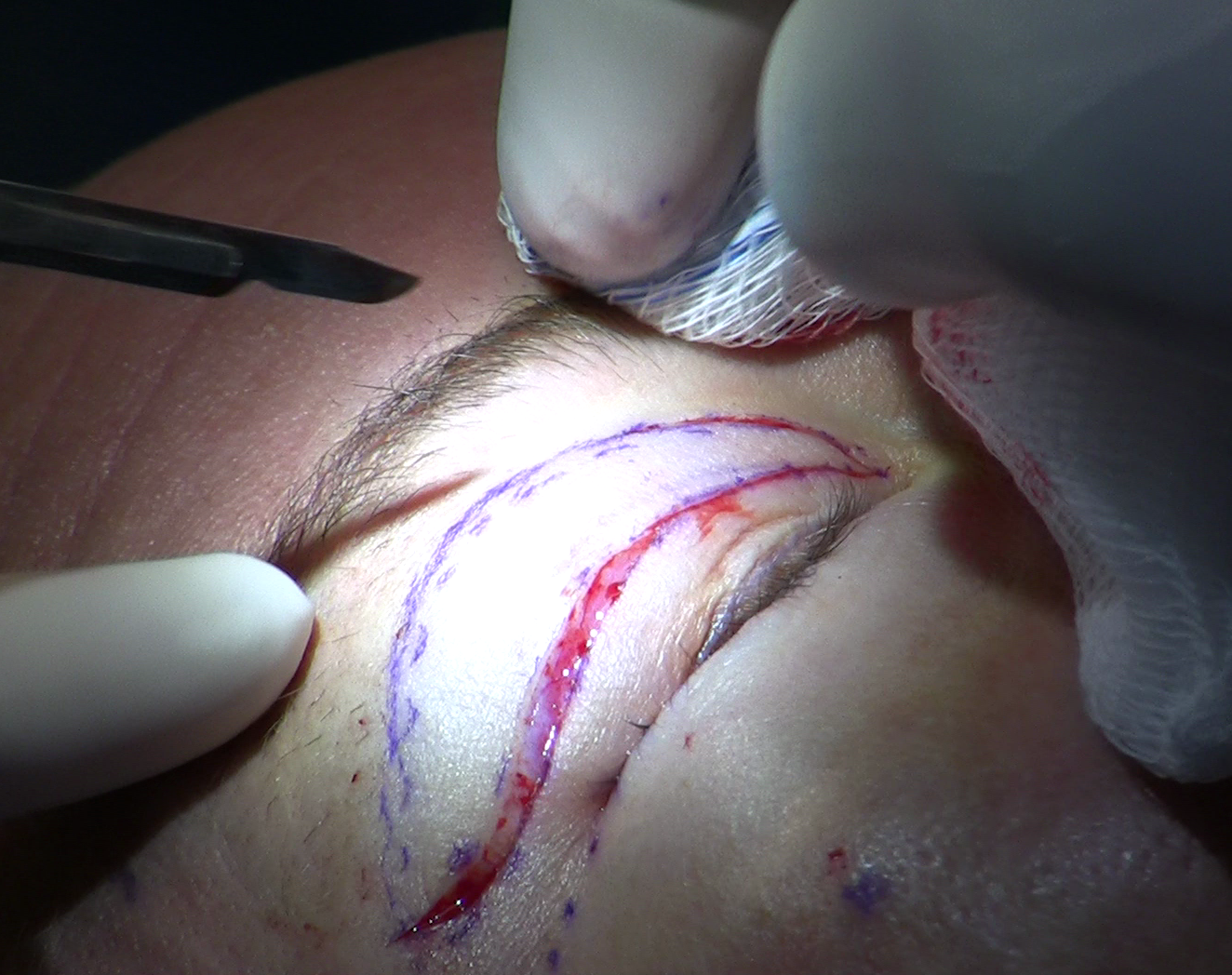
What Retraction Does to the Eye
The cornea depends on a complete blink to re-spread the tear film. When the upper lid sits too high, each blink is incomplete - the lid travels but does not fully cover the inferior and central cornea. Those zones dry between cycles, and over time the corneal epithelium breaks down into fine surface erosions called superficial punctate keratitis. The result is pain, light sensitivity, and blurred vision from a roughened corneal surface.
Untreated lagophthalmos exposes the cornea for hours without any protective blink. This can progress from punctate erosions to frank corneal ulceration, with risk of permanent scarring and vision loss in severe cases. Separately, the staring appearance of upper lid retraction carries a real quality-of-life cost - patients consistently describe being told they look surprised or aggressive when they feel entirely composed.
Diagnosis and Measurement
The standard measurement is MRD1 - margin-to-reflex distance. The examiner holds a penlight directly in front of the patient's eye and measures from the upper lid margin to the corneal light reflex. A normal upper lid MRD1 is 4-5 mm. Retraction is diagnosed when MRD1 exceeds 5.5 mm, or when the difference between the two eyes is greater than 1 mm - because asymmetry alone is an abnormal finding even if neither eye crosses the absolute threshold individually.
A complete evaluation follows a logical sequence:
- Measure MRD1 in both eyes in primary gaze, and assess for lid lag in down-gaze (a sign of thyroid-related fibrosis where the lid lags behind the globe on downward movement)
- Examine the cornea with fluorescein dye and a slit lamp to detect existing punctate erosions or frank exposure damage before any treatment begins
- Perform Schirmer's test to establish baseline tear production - patients with already-compromised tear production face greater exposure risk and need more aggressive lubrication
- Order thyroid function tests (TSH, free T4, thyroid antibodies) to identify or confirm thyroid eye disease
- Obtain orbital CT or MRI when thyroid disease is suspected - enlarged extraocular muscles and fat expansion are diagnostic, and imaging guides surgical planning for decompression if needed
- Document lid position photographically in standardized conditions, creating a baseline for tracking change and for planning surgical endpoints
Non-Surgical Management and the Waiting Period
For many patients, the first phase of care is protective rather than corrective. The goal is preventing corneal damage while the cause is evaluated and treated, or while waiting for thyroid eye disease to stabilize.
- Preservative-free lubricating drops used frequently through waking hours - the preservatives in standard drops can irritate a compromised corneal surface with repeated use
- Viscous gel drops for more sustained corneal coverage during the day
- Lubricating ointment applied at the lid margin at night, before sleep
- Moisture-chamber goggles worn overnight to maintain humidity around the eye
- Bandage contact lenses, placed by the ophthalmologist, for corneas with active epithelial breakdown that needs a physical barrier while healing
The 6-month stability rule for thyroid eye disease is not arbitrary caution. The active inflammatory phase actively remodels lid tissue - a lid position achieved surgically in that phase may continue to change as fibrosis progresses, erasing the correction. Some patients see spontaneous improvement in retraction as the active phase resolves, reducing the degree of surgical correction needed later.
Teprotumumab (Tepezza), an IGF-1 receptor inhibitor approved in 2020, is administered during the active phase of thyroid eye disease to reduce orbital inflammation. It decreases proptosis and, in a meaningful proportion of patients, reduces eyelid retraction - in some cases enough to avoid surgery or make surgical correction more straightforward. It is given as an infusion series under physician supervision. It does not replace surgical correction once the disease is inactive and retraction persists, but it can meaningfully change what surgery is ultimately needed.
The Three Surgical Techniques
When retraction persists after stability is confirmed and lubrication is insufficient to protect the cornea, surgery addresses the shortened or overactive elevator tissue. Three approaches are used, selected by the degree of retraction and the amount of tissue available to recess.
| Technique | Typical Severity | Surgical Access | Spacer Required |
|---|---|---|---|
| Müller muscle recession | Mild (approx. 1-2 mm above normal) | Posterior - transconjunctival, no skin incision | Rarely |
| Levator recession combined with Müller recession | Moderate to severe (greater than 2 mm) | Anterior through skin crease, or combined | Sometimes |
| Spacer graft with recession | Severe or insufficient native tissue | Anterior - full lid exposure | Always - the graft is the core of the repair |
Müller Muscle Recession
For mild retraction, recessing the Müller muscle alone through a posterior (transconjunctival) approach is frequently adequate. The surgeon enters from the inside surface of the lid, through the conjunctiva, with no skin incision at all. This is particularly important for patients who have had prior anterior lid surgery - blepharoplasty, ptosis repair, or orbital work. Reopening old scar tissue externally risks adding new scarring to an already-compromised lid. The posterior route avoids that plane entirely and leaves no visible incision.
Levator Aponeurosis Recession
Greater degrees of retraction require recessing the levator aponeurosis itself - moving it posteriorly to reduce its mechanical pull on the tarsus. In a published American Journal of Ophthalmology series, combined Müller plus levator recession lowered mean MRD1 from 6.0 mm to 3.4 mm, a correction of 2.6 mm, with a failure rate of only 8.4%. Most of those under- or over-corrections were successfully revised with a second procedure. These figures describe a reproducible, well-characterized operation when performed by surgeons experienced with the anatomy.
Spacer Grafting
After recession, the surgeon sometimes finds insufficient tissue to hold the lid in its new lower position without undue tension. A spacer graft fills the gap - tissue interposed between the recessed edges to add length. Autologous options include hard palate mucosa (durable, but requires a second surgical site inside the mouth with some temporary donor-site soreness) and free tarsal grafts from the fellow eyelid (excellent tissue match, minimal donor morbidity). Allograft materials - including acellular dermal matrix - avoid any donor site and are available immediately, though long-term data for these materials is more limited. Donor sclera, once used commonly, is increasingly avoided because it can shrink progressively over time.
Outcomes, Adjustable Sutures, and Choosing the Right Surgeon
In a 12-year study of Müller's muscle excision combined with levator recession, 96% of treated lids achieved a position within 1.5 mm of the fellow eyelid, and mean postoperative asymmetry improved from 1.0 mm to 0.4 mm - well within the range that registers as symmetric to an observer.PubMed PMID 3755520
Adjustable sutures allow the surgeon to set the recession without permanently tying the knots at the time of surgery. In the first 24-48 hours after the procedure, while the patient is awake and the local anesthetic has worn off, the surgeon examines the lid position and fine-tunes suture tension under topical anesthesia. The patient can cooperate with the exam, the lid can be observed in natural lighting and gaze positions, and the final height is confirmed before the tissue heals into place. This technique specifically addresses one of the main risks of lid-height surgery - residual asymmetry from the unpredictability of individual healing.
Risks across all three approaches include undercorrection, overcorrection into ptosis, asymmetry between the two lids, and dry eye exacerbation. Upper and lower lid retraction frequently coexist in thyroid eye disease - the upper lid pulls up and the lower lid pulls down, exposing the cornea from both directions. Each requires its own repair, and they are typically staged. Patients who also need orbital decompression or strabismus surgery follow a specific sequence: decompression first, then muscle surgery, then lid repair, because each alters the anatomical context for the next.
Frequently Asked Questions
How would I know if my upper lid is retracted rather than just naturally wide?
The clinical marker is visible white sclera between the upper lid margin and the iris - a zone that should be hidden under the lid. True retraction also produces a measurable MRD1 above the normal range, or measurable asymmetry between the two eyes. An oculoplastic surgeon can take this measurement at a routine office visit and compare it to baseline photographs if available. Natural lid width varies, but visible superior scleral show is not a normal variant.
If I had ptosis surgery and now my other eye looks too wide, was something done wrong?
Not necessarily. This is Hering's Law operating as expected: before your ptosis was repaired, the shared levator drive was elevated in both eyes to compensate, which was silently holding the fellow lid high. After the correction drops that drive, the fellow lid reveals retraction that was already present but masked. A thorough preoperative evaluation for ptosis surgery includes looking for this pattern in the fellow lid specifically, so the possibility can be discussed before the first operation rather than after.
Can retraction improve without surgery in thyroid eye disease?
Partial spontaneous improvement does occur as the active phase of thyroid eye disease resolves - inflammation subsides, and the fibrotic pull on the lid can soften somewhat. Teprotumumab, given during the active phase, has produced measurable reduction in lid retraction in a portion of patients. Surgery is reserved for retraction that persists after the disease has been inactive for at least 6 months, because operating before stability is established produces unreliable results as the underlying tissue continues to change.
What is a spacer graft and why would I need one?
After the elevator muscles are recessed, the surgeon sometimes finds that the recessed edges cannot hold the lid at its new, lower position without pulling the lid back up - there is simply not enough tissue length to span the gap comfortably. A spacer graft fills that gap with an additional piece of tissue. The material can be your own hard palate lining, a small piece of tarsus from the opposite lid, or an allograft material that does not require a donor site. Which one is used depends on how much length is needed and your individual anatomy - your surgeon will discuss the options specific to your case.
How long does it take to see the final result after retraction surgery?
Most visible swelling resolves within 2-3 weeks, and patients generally return to office work within 1-2 weeks. Final lid position, however, continues to settle as scar tissue matures - the full result may not be apparent for up to 3 months. Adjustable sutures, when used, are fine-tuned in the first day or two post-operatively to set the starting height accurately. Lubricating drops are continued throughout recovery and often indefinitely for patients with underlying dry eye from thyroid disease or prior surgery.
General information only, not medical advice. Individual anatomy and healing vary. See the disclaimer.