Most chalazions resolve after a single incision-and-curettage and never return. Sebaceous carcinoma does not work that way. It grows inside the eyelid for months - sometimes years - producing a bump that looks and behaves exactly like a blocked gland while it spreads through tissue that shows no visible sign of involvement. By the time pathology confirms the diagnosis, many patients have already had two or three unsuccessful procedures. Understanding why this cancer hides so effectively, and what a complete diagnostic and treatment sequence actually requires, is the difference between early cure and late-stage disease.
What Sebaceous Carcinoma Is - and Why the Upper Eyelid Bears the Burden
Sebaceous carcinoma arises from oil-producing glands, and the eyelid contains three populations of them. Meibomian glands run vertically through the tarsal plate of each lid, opening at the lid margin with dozens of secretory orifices. Glands of Zeis are smaller sebaceous units attached to individual eyelash follicles. The caruncle, the pink nodule at the inner corner of the eye, contains modified sebaceous tissue as well. Any of these three sources can produce this cancer, but meibomian glands account for the large majority of cases.
The anatomy of the tarsal plate directly explains one of the most consistent findings in published case series: roughly 63% of sebaceous carcinomas originate in the upper eyelid, compared to about 27% in the lower eyelid, with the remaining cases involving both lids simultaneously. The upper tarsus contains approximately twice as many meibomian glands as the lower tarsus. More secretory glandular tissue means more cells available for malignant transformation, and the roughly 2:1 gland-density ratio tracks almost exactly with the two-to-three times higher cancer incidence in the upper lid.
Despite this anatomy, sebaceous carcinoma remains uncommon overall - it accounts for less than 1% of all eyelid tumors and ranks third among eyelid malignancies, behind basal cell and squamous cell carcinoma. The challenge is not frequency; it is behavior. No other eyelid cancer spreads the way sebaceous carcinoma does, and no other eyelid cancer mimics benign disease as consistently.
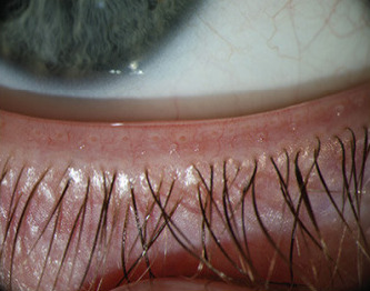
The Masquerade Syndrome: Four Diagnoses This Cancer Impersonates
The term "masquerade syndrome" describes a recognized clinical pattern, not a metaphor. Sebaceous carcinoma produces signs that are clinically indistinguishable from four common benign conditions:
- Chalazion - a firm lid nodule from a blocked meibomian gland, by far the most frequent misdiagnosis
- Diffuse blepharitis - lid-margin inflammation with redness, scaling, and crusting
- Chronic conjunctivitis - persistent eye redness and discharge that fails to resolve with standard treatment
- Superior limbic keratoconjunctivitis - inflammation at the upper limbus where the cornea meets the white of the eye
The published median time from first symptoms to correct histopathologic diagnosis is 12 months, with a range extending to 60 months. One review of over a thousand specimens clinically diagnosed as chalazion found a misdiagnosis rate of 6.4%; of the missed malignancies in that cohort, 80% were sebaceous carcinoma. These are not extreme statistical outliers - they reflect a recurring failure of pattern recognition that occurs in academic centers as well as community practices.
A chalazion that recurs after incision-and-curettage, involves the upper lid, and is accompanied by lash loss or unilateral lid thickening warrants biopsy before any further procedural treatment - not another I&C.
The following signs should shift clinical thinking toward biopsy rather than repeat treatment:
- Rapid recurrence of a chalazion in the same location after prior drainage
- Loss of eyelashes (madarosis) adjacent to the nodule
- Unilateral blepharitis that does not respond to lid hygiene and topical antibiotics
- Firmness or thickening of the tarsal plate beyond what a simple cyst explains
- Persistent conjunctival injection without identifiable infectious cause
- Any chalazion-like lesion in a patient with prior head or neck radiation
Who Is at Risk
Sebaceous carcinoma is primarily a disease of older adults, with peak incidence after age 60. Women are affected more often than men, and Asian patients have a documented higher incidence than other racial groups. Beyond these baseline demographics, several specific exposures increase risk substantially:
- Prior radiation to the head or neck - the cancer can appear 20 to 60 years after exposure, making radiation history clinically relevant even in patients treated decades earlier
- Immunosuppression from any source, including organ transplantation, HIV infection, or prolonged immunosuppressive therapy
- Muir-Torre syndrome, a heritable cancer syndrome addressed in a later section - patients under 60 with this diagnosis deserve particular scrutiny for the genetic connection
Getting the Diagnosis Right: The Procedure Matters as Much as the Pathologist
When sebaceous carcinoma is on the differential, the biopsy technique determines whether the specimen is interpretable. Incision-and-curettage - correct procedure for a chalazion - crushes and fragments tissue. The diagnostic hallmarks of sebaceous carcinoma are intracytoplasmic lipid vacuoles and a specific glandular architecture that must arrive at the pathology lab intact.
A full-thickness eyelid biopsy - a small wedge that includes skin, orbicularis muscle, tarsus, and conjunctiva in continuity - preserves that architecture. When standard hematoxylin-and-eosin staining leaves the diagnosis uncertain, immunohistochemistry resolves the ambiguity. Sebaceous carcinoma stains positively for androgen receptor, EMA, and adipophilin, which marks the intracellular lipid characteristic of sebaceous differentiation. Basal cell carcinoma and squamous cell carcinoma do not share this profile, allowing confident differentiation even in poorly differentiated tumors where cell morphology overlaps.
Every sebaceous carcinoma specimen should also undergo mismatch repair protein immunohistochemistry at the time of initial diagnosis - not as an afterthought, but as part of the standard workup. The reason is explained in the Muir-Torre section.
Pagetoid Spread and Conjunctival Map Biopsies
Pagetoid spread is the feature that makes sebaceous carcinoma more surgically complex than any other eyelid malignancy. Tumor cells migrate laterally through the conjunctival epithelium - the thin membrane lining the inside of the lids and the front of the eye - without forming a visible surface mass. This spread is invisible on slit lamp examination. The conjunctiva looks completely normal while malignant cells are moving through it.
This pattern is unique to sebaceous carcinoma among eyelid cancers. A surgeon who excises the visible tumor without first mapping pagetoid extent will leave tumor cells behind in tissue that appeared clear, and the cancer will recur.
Conjunctival map biopsies address this problem directly. Before any definitive excision, the surgeon takes 10 to 14 small conjunctival specimens from standardized locations - multiple sites from the tarsal conjunctiva lining each lid, multiple sites from the bulbar conjunctiva over the globe, and specimens from the limbus. Each specimen is labeled by site and sent separately to pathology. The results produce a spatial map of where tumor cells are present, which defines the true surgical boundaries before the main operation begins.
Surgical Treatment, Topical MMC, and Reconstruction
Surgery is the only reliable curative treatment. Published data show a local recurrence rate of approximately 71% when radiation serves as the primary treatment, compared to roughly 22% with surgical excision - a difference large enough that radiation alone is not a clinically acceptable substitute. The full treatment sequence proceeds in stages over days to weeks:
- Full-thickness biopsy confirms the diagnosis histopathologically.
- Conjunctival map biopsies define pagetoid extent before any excision begins.
- Mohs micrographic surgery or CCPDMA (complete circumferential peripheral and deep margin assessment) removes the primary tumor while examining 100% of the surgical margin - staged, layer by layer, until all margins are clear.
- If map biopsies showed conjunctival pagetoid involvement, topical mitomycin C is applied to the ocular surface after excision. Published series using MMC in alternating weekly on-off cycles report complete resolution of pagetoid disease in most patients, sparing the eye from more radical surgery.
- Sentinel lymph node biopsy is performed as indicated by staging.
- Lid reconstruction follows once clear margins are confirmed.
For large upper-eyelid defects - the most common scenario given upper-lid predominance - reconstruction options include Cutler-Beard advancement flaps from the lower lid, Hughes tarsoconjunctival flaps combined with skin grafts, and temporal or forehead flaps for extensive external surface defects. These are staged procedures; most require a second operation several weeks later to separate the tissue bridge and fully restore lid function.
AJCC Staging and Sentinel Lymph Node Biopsy
AJCC eyelid tumor staging classifies tumors by size and local extent. T1 describes a small tumor confined to the lid; T2 indicates larger size or early involvement of adjacent structures; T3 reflects orbital extension; T4 denotes very advanced local disease. Sentinel lymph node biopsy - sampling the first draining lymph node from the tumor site - is recommended at T2c staging and above. It is also triggered regardless of T-stage by any of these findings:
- Pagetoid spread confirmed on map biopsies
- Perineural invasion on histopathology
- Poor differentiation
- Active immunosuppression
A positive sentinel node - meaning tumor cells detected in regional lymph nodes - significantly changes prognosis and triggers discussion of systemic therapy or additional regional treatment. At that point the disease is no longer confined to the eyelid, and the surgical cure rate alone is lower.
Muir-Torre Syndrome and Genetic Implications for the Whole Family
Muir-Torre syndrome is a hereditary cancer syndrome caused by germline mutations in mismatch repair genes, specifically MLH1 and MSH2. It is a variant of Lynch syndrome - the same underlying genetic mechanism that predisposes families to colorectal, endometrial, and other visceral cancers. In Muir-Torre syndrome, the same mutation also produces sebaceous tumors of the skin and eyelid.
The critical clinical point: in some patients, the eyelid sebaceous carcinoma appears before any internal malignancy has been diagnosed. The eyelid tumor is the first warning that a Lynch-spectrum cancer may be developing elsewhere.
Screening begins at the pathology lab. MMR protein immunohistochemistry on the biopsy specimen tests for loss of MLH1, MSH2, MSH6, or PMS2 protein expression. Absent expression signals a deficient repair system and warrants genetic counseling and germline testing. The screening threshold should be lower in patients diagnosed before age 60.
A confirmed germline mutation carries consequences beyond the patient's own surveillance plan. First-degree relatives have a substantial probability of carrying the same mutation and should be offered testing. Those who carry it need a dedicated surveillance program - colonoscopy, gynecologic screening, and cancer surveillance matched to their specific mutation. A single eyelid biopsy, interpreted correctly, can redirect cancer prevention for an entire family.
Prognosis, Surveillance, and Long-Term Outlook
The table below places sebaceous carcinoma alongside the eyelid cancers most patients are more likely to encounter.
| Feature | Basal Cell Carcinoma | Squamous Cell Carcinoma | Sebaceous Carcinoma |
|---|---|---|---|
| Most common lid site | Lower lid / medial canthus | Lower lid | Upper lid |
| Pagetoid conjunctival spread | No | No | Yes - unique to this cancer |
| Map biopsies required | No | No | Yes, before definitive excision |
| Radiation as primary treatment | Accepted option | Sometimes used | High recurrence - surgery required |
| Inherited cancer syndrome link | Gorlin syndrome (rare) | Not typically | Muir-Torre / Lynch syndrome |
Five-year disease-specific survival for localized sebaceous carcinoma is approximately 94%, which reflects how well surgery performs when the diagnosis is made before spread has occurred. Once regional lymph nodes are involved, that figure falls sharply - a gap that underscores why the 12-month diagnostic delay documented in published literature is not merely an inconvenience.
Local recurrence after excision occurs in 9 to 36% of cases; regional metastasis in 3 to 25%. Most recurrences develop within the first five years, which defines the minimum meaningful follow-up window. Three independent factors predict worse outcomes: a diagnostic delay longer than six months, a tumor diameter greater than 1 cm at the time of surgery, and simultaneous involvement of both upper and lower eyelids at presentation.
Follow-up examinations typically occur every three to six months for the first two years, then annually. Each visit should include inspection of both lids, the conjunctival surface, and palpation of regional lymph nodes - not just visual review of the surgical scar. Any new firmness, nodule, or conjunctival irregularity warrants prompt biopsy.
Orbital exenteration - removal of the entire orbital contents - was historically used for advanced disease. The combination of thorough map biopsies, staged margin-controlled excision, and topical MMC for pagetoid disease has substantially reduced the circumstances in which that degree of surgery is necessary. Exenteration is now reserved for documented orbital invasion that cannot be cleared by less radical approaches.
Frequently Asked Questions
How do I know if my chalazion should be biopsied rather than just treated again?
Any chalazion that returns after incision-and-curettage in the same location warrants a biopsy before a repeat procedure. Additional reasons to biopsy include lash loss near the nodule, firmness or thickening of the tarsal plate that extends beyond a discrete cyst, upper-eyelid location in a patient over 60, and any prior history of head or neck radiation. A recurrent chalazion that does not respond to treatment is a different clinical problem from a first-time blocked gland.
Why does pagetoid spread change the surgical plan so significantly?
Because the surgeon cannot see it. Tumor cells can be migrating through the conjunctiva across a wide area while the tissue looks completely normal to the naked eye and under a slit lamp. Without map biopsies taken before the main operation, the surgeon has no way to know where the true margins of disease are, and excision based only on the visible tumor will leave cells behind. The map biopsy results tell the surgeon - and the patient - what they are actually dealing with before reconstruction is planned.
Is sebaceous carcinoma connected to inherited cancer syndromes?
Yes. Muir-Torre syndrome, a variant of Lynch syndrome, is caused by inherited mutations in mismatch repair genes and predisposes carriers to both sebaceous tumors and internal cancers including colorectal and endometrial cancer. Pathologists can screen for this on the initial biopsy using standard protein staining. A positive result should lead to genetic counseling, and first-degree relatives should be offered testing because they have a meaningful probability of carrying the same mutation and needing their own surveillance program.
Why is radiation not used the way it is for basal cell carcinoma of the eyelid?
Published outcomes data show a local recurrence rate of approximately 71% when radiation serves as the primary treatment for sebaceous carcinoma, compared to roughly 22% with surgical excision. That gap is too large to accept as a primary strategy. Radiation may have a role as an adjunct after surgery in specific circumstances, but it does not replace complete surgical excision for this particular cancer.
What does the follow-up schedule look like after surgery?
Most patients are seen every three to six months for the first two years, then annually thereafter. Every visit should include inspection of the surgical site, both lids, the conjunctival surface, and regional lymph nodes - because recurrence can appear at any of those sites, not only where the original tumor was. The five-year window is the highest-risk period, but annual surveillance beyond that remains appropriate given this cancer's known behavior.
Does having sebaceous carcinoma mean my children need cancer screening too?
It depends on whether your tumor shows loss of mismatch repair protein expression on immunohistochemistry. If it does, and genetic testing confirms a germline mutation, first-degree relatives have a significant chance of carrying the same alteration and should be offered genetic counseling and testing. Those who carry it will need a structured surveillance program for Lynch-associated cancers. Your treating oculoplastic surgeon can coordinate a referral to a genetic counselor if the MMR staining result warrants it.
General information only, not medical advice. Individual anatomy and healing vary. See the disclaimer.