When a surgeon recommends browpexy alongside upper eyelid surgery, the suggestion typically follows a specific observation: the outer third of your brow has descended far enough to push excess skin onto the upper lid, and correcting the lid skin alone - without addressing that descent - would either leave the result incomplete or require removing more skin than is safe. Browpexy is a brow-stabilization procedure performed entirely through the eyelid crease incision already planned for blepharoplasty. No separate forehead or hairline cut is required. Its appeal is real, but so are its limits. Understanding both starts with the tissue mechanics that make the outer brow the most vulnerable part of the brow complex to begin with.
Why the Lateral Brow Falls First: Periosteal Attachment Anatomy and the ROOF Fat Pad
The brow is not attached uniformly to the bone beneath it. Its periosteal connections - the fibrous bands that anchor soft tissue to the frontal and supraorbital bone - are substantially firmer over the medial half to two-thirds of the orbit. Toward the outer corner, those attachments weaken progressively. This structural imbalance means that as facial ligaments relax over time, the lateral brow descends first and most noticeably, while the medial brow can hold its position for years or even decades longer. The characteristic outer-brow droop of middle age is not random - it is the direct consequence of where the periosteal tethering is thin.
Sitting between the orbicularis oculi muscle and the periosteum of the frontal bone is a distinct fibrofatty layer called the retro-orbicularis oculi fat, abbreviated ROOF. This pad is not passive tissue. It is the primary gliding plane the brow traverses during facial expression - the layer that allows the brow to move upward and downward without being tethered to the skull. The ROOF sits superior to the orbital septum, directly above the supraorbital rim. In youth, it contributes to the fullness and slight projection of the lateral brow. With age, the ROOF deflates and the overlying tissues lose structural support, accelerating lateral brow descent even before visible skin laxity appears in the forehead.
The ROOF is also the operative space surgeons enter during browpexy. The sub-ROOF plane - the layer between the ROOF and the underlying periosteum - provides the dissection corridor that allows a suture placed through a blepharoplasty incision to reach and anchor to bone without a separate forehead approach.
What Browpexy Is - and Where It Sits Between Upper Blepharoplasty and a Full Brow Lift
Browpexy occupies the middle ground between two distinct operations. Upper blepharoplasty addresses the upper eyelid skin and sometimes the underlying orbital fat compartments; it does nothing to the brow itself. A formal brow lift - endoscopic, direct, or coronal - repositions the brow through forehead or hairline dissection with significant tissue release and fixation. Browpexy is neither. It is a fixation-only procedure that stabilizes the lateral brow in a slightly elevated position without requiring the forehead dissection a true brow lift demands.
The procedure adds approximately ten to twenty minutes to a blepharoplasty and produces no separate visible scar. The trade-off is that the correction is modest. Browpexy cannot achieve the elevation of a formal brow lift, cannot address forehead skin or the medial brow, and cannot substitute for a brow lift when the degree of descent is beyond what a suture placed through the eyelid can adequately resolve. Knowing where that line falls is the most important judgment a surgeon makes when planning combined eyelid and brow work.
Internal (Transblepharoplasty) Browpexy: What the Surgeon Actually Does, Step by Step
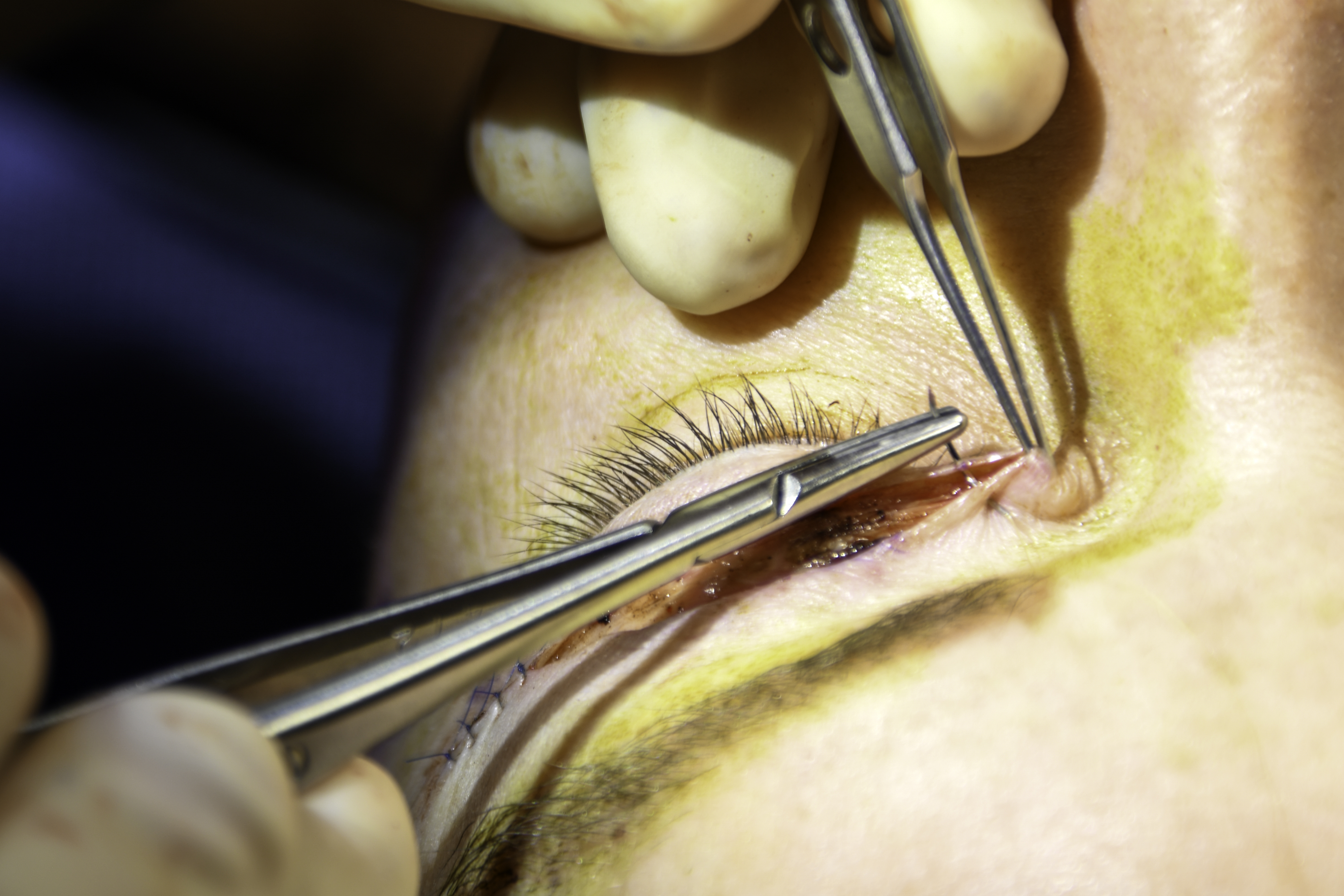
The internal technique - sometimes called the BUL (Browpexy through the Upper Lid) technique after its formal description in the Aesthetic Surgery Journal in 2011 - proceeds through the same incision already made at the upper eyelid crease. The sequence below describes the operative approach.
- Crease incision and initial dissection. The surgeon opens the upper eyelid crease incision and completes the blepharoplasty portion of the work - addressing skin, orbicularis, and orbital fat as planned.
- Superior dissection toward the orbital rim. The surgeon extends the dissection upward through the post-orbicularis plane, separating the orbicularis oculi from the underlying ROOF and then entering the sub-ROOF plane directly above the periosteum of the frontal bone.
- Periosteal identification under direct vision. Working through the sub-ROOF corridor, the surgeon exposes the frontal bone periosteum above the supraorbital rim. The supraorbital neurovascular bundle - which runs medially along the mid-supraorbital rim inside the frontalis - is identified and avoided; the fixation point is placed well above the orbital rim and lateral to this structure.
- Suture placement through orbicularis or brow fat pad. An absorbable suture - most commonly 4-0 polyglactin 910 (Vicryl) - is passed through the orbicularis oculi or the brow fat pad and then anchored to the periosteum at the fixation point. In the 'brassiere suture' variant, the orbicularis itself is sutured directly to the periosteum of the superior lateral orbital rim, named for the geometry of the suspension.
- Tension assessment and symmetry check. Before the suture is tied, the surgeon assesses brow position and symmetry under gentle tension. Over-elevation is actively avoided because brow position cannot easily be adjusted downward after final fixation.
- Closure. The eyelid crease incision is closed in the standard blepharoplasty fashion. No additional wound is present because no additional incision was made.
The Vicryl suture used in internal browpexy begins losing tensile strength progressively from the first week, retaining approximately 75% of original strength at two weeks and approximately 50% at three weeks, and fully resorbs by roughly eight to ten weeks. It does not provide permanent fixation - it holds the tissues in place long enough for fibrosis to form between the ROOF and the periosteum at the anchor site. That scar tissue becomes the durable mechanical support. If the fibrosis is well-formed, position is maintained; if soft-tissue healing is incomplete before the suture dissolves, the brow can gradually settle back toward its preoperative level.
External Browpexy: The Brow-Hair Incision Approach and How Its Fixation Differs
External browpexy uses the same general principle - suture fixation of brow soft tissue to frontal bone periosteum - but approaches from above rather than below. A small incision, hidden within the brow hairs at the lateral brow, gives the surgeon direct access to the orbicularis and brow fat pad, which are then anchored to the periosteum through this upper approach.
The critical difference is suture type. External browpexy typically employs a permanent non-absorbable suture. Because the fixation does not depend on a window of fibrosis formation before the suture dissolves, the mechanical hold is more durable from the outset. A long-term comparison study published around 2020 found that internal and external browpexy combined with blepharoplasty produced comparable lateral brow elevation at 24 months, with any short-term advantage for external browpexy attributed to permanent suture fixation rather than technique alone; the relative durability difference between techniques remains an active area of discussion in the literature. The trade-off is the additional incision within the brow hairs, which in patients with sparse lateral brows may be more noticeable over time. For patients who prioritize longevity of correction over avoiding a second wound, the external approach makes a stronger mechanical case - confirm with your surgeon what the current evidence shows for your specific anatomy.
The Skin-Excision Problem: Why Adding Browpexy Forces a More Conservative Blepharoplasty Plan
This interaction between browpexy and blepharoplasty is the most clinically significant aspect of the combined procedure - and it is almost never explained to patients before surgery.
When the brow is elevated, even modestly, it mechanically stretches the upper eyelid skin upward. Skin that appeared redundant at the preoperative consultation will be lifted by the brow correction and appear less redundant after fixation. If a surgeon marks the planned skin excision based on the brow's pre-correction position and then adds browpexy during the same operation, the amount of skin removed may exceed what is now safe given the elevated brow. The result, in the worst case, is lagophthalmos - the inability to fully close the eyelid - which leaves the cornea exposed and unprotected. Lagophthalmos after eyelid surgery is not a minor inconvenience; it can cause corneal drying, surface damage, and significant discomfort.
To avoid this, surgeons performing combined browpexy and upper blepharoplasty apply a more conservative excision plan - reducing the planned skin removal by a small margin to account for the skin stretch the brow correction will produce. Some surgeons prefer to perform the browpexy fixation first, assess the new lid geometry with the brow held in its corrected position, and then mark the skin excision accordingly. Either approach requires that the interaction be explicitly anticipated before cutting, not discovered after.
How Much Lift to Expect and How Long Results Last: What the Data Show
Patients are routinely told browpexy 'gently lifts' the brow without a concrete frame of reference for what that means. Published outcome data allow a more specific description.
A 2018 study (Zandi et al., Journal of Cosmetic Dermatology) measured lateral brow elevation in women undergoing internal browpexy with upper blepharoplasty and found a modest but anatomically meaningful improvement at six months compared to preoperative baseline, concentrated at the outer brow. The specific millimeter value cited in some secondary sources could not be independently confirmed from available abstracts; consult the primary source for the exact reported figure.Zandi et al., Journal of Cosmetic Dermatology, 2018
For context, a 2025 systematic review and meta-analysis of endoscopic brow lift outcomes found a pooled lateral brow elevation of approximately 4.3 mm - roughly double or more what browpexy typically produces - and the endoscopic lift also addresses medial brow position, forehead skin, and glabellar creases, none of which browpexy changes.
Long-term durability varies with technique, tissue quality, and individual healing. Internal browpexy relies on fibrosis at an absorbable suture site; that scar tissue can soften and stretch over years, allowing gradual re-descent. External browpexy's permanent suture provides a longer-lasting mechanical anchor, though the surrounding soft tissues continue to age around it. Neither technique permanently arrests brow descent.
| Technique | Incision | Suture type | Published lateral lift | Relative durability |
|---|---|---|---|---|
| Internal browpexy | Eyelid crease only | Absorbable (Vicryl) | Approx. 2-3 mm at 6 months (verify with primary sources) | Moderate - fibrosis-dependent |
| External browpexy | Eyelid crease + small brow incision | Permanent non-absorbable | Similar to internal; long-term results comparable per published data | Moderate to greater - permanent anchor, though long-term comparisons show similar outcomes to internal at 24 months |
| Endoscopic brow lift | Scalp ports, no visible face scar | Various fixation methods | Approx. 4-5 mm pooled (2025 meta-analysis) | Greater - full periosteal release |
| Direct brow lift | Above brow or in forehead crease | Permanent or absorbable | Surgeon-controlled; higher ceiling | High - direct soft tissue excision |
Who Is the Right Candidate - and the Clear Signs That a Full Brow Lift Is the Better Answer
Browpexy delivers its best results for a specific patient profile. Outside that profile, performing browpexy instead of a formal brow lift exchanges a small additional scar for a result that does not adequately solve the underlying problem.
The ideal browpexy candidate typically has most of the following characteristics:
- Mild to moderate lateral brow descent, with the medial brow sitting in an acceptable position
- Upper eyelid skin redundancy that is at least partly driven by lateral brow depression, not lid skin excess alone
- No significant excess forehead skin - the forehead itself is in an acceptable position
- Realistic expectations of a modest repositioning effect, not a dramatic change in brow height
- A preference to avoid a separate forehead or hairline incision and its associated recovery
The following findings indicate that a formal brow lift is the appropriate operation, and that browpexy will likely underperform:
- Severe or global brow ptosis. When the entire brow has descended substantially - not just the outer third - suture fixation through the eyelid cannot restore the brow to an appropriate position. The degree of elevation required exceeds what a transblepharoplasty suture can deliver.
- Medial-predominant brow descent. The suture fixation point in internal browpexy must stay well lateral to the supraorbital nerve. Browpexy cannot effectively address a brow whose primary descent is at its medial end. A direct or endoscopic approach is required to correct medial brow ptosis safely.
- Significant forehead skin laxity. When the forehead itself carries excess skin, brow fixation does not address the fundamental problem. A procedure that repositions or removes forehead tissue is the appropriate solution.
- Post-facial palsy brow asymmetry. Paralytic brow ptosis behaves differently from age-related descent. The underlying muscle dysfunction means suture fixation is unlikely to produce symmetric, lasting results, and formal procedures with greater tissue control are required.
- Prior failed browpexy. When browpexy has already been performed and failed to maintain correction, repeating it rarely achieves better results. The fixation site has already scarred, and the tissues have demonstrated their tendency to re-descend.
Recovery: What the Browpexy Healing Timeline Looks Like When Combined With Eyelid Surgery
Adding browpexy to upper blepharoplasty does not meaningfully extend the postoperative recovery compared to blepharoplasty alone. The procedure adds time in the operating room but confines the additional tissue disruption to the eyelid crease region already involved in the blepharoplasty, with the superior dissection into the sub-ROOF plane producing only modest additional swelling above the brow.
The early recovery follows the standard blepharoplasty course. Bruising and swelling peak in the first two to three days. The brow area may feel firm or mildly elevated in the first week as swelling fills the dissection space; this settles as healing progresses. Most patients are comfortable in public settings within two weeks.
Because internal browpexy depends on fibrosis forming at the suture anchor site before the Vicryl resorbs, the first six to ten weeks represent a critical window. During this period, patients should avoid the following:
- Aggressive massage or manipulation of the brow or lateral forehead
- Contact sports or activities that risk direct impact to the brow area
- Elective procedures involving the forehead - lasers, injectables near the fixation site - without the surgeon's clearance
- Activities that substantially increase facial tissue tension before the anchor site has matured
By the time the suture has fully resorbed, fibrosis at the fixation site should be established. Brow position at three months is generally representative of what the long-term result will be, though continued soft-tissue aging will gradually affect position over years regardless of initial outcome.
Billing and Insurance: Why Browpexy Must Be Documented Separately Even in Functional Cases
Functional upper blepharoplasty - performed because eyelid skin is obstructing the visual field - can qualify for insurance coverage with appropriate documentation, including visual field testing and clinical photography. Browpexy does not qualify under any circumstances.
Even when browpexy is performed at the same time, through the same incision, and in the same operative session as a covered functional blepharoplasty, insurers classify brow elevation as a cosmetic service. The operative note must separately document the browpexy as a cosmetic procedure with separate coding and a separate patient financial responsibility. Combining the two services into a single operative description, or omitting the browpexy from the documentation, creates billing errors with insurance and compliance implications for the practice.
This distinction matters practically. Patients who assume that insurance coverage for functional blepharoplasty extends to all work performed in that session may be surprised by out-of-pocket costs for the browpexy. Clarifying what will be submitted to insurance versus billed directly - and getting that breakdown in writing - should happen before the procedure date, not at discharge. The surgeon's billing staff can provide documentation of each service's coding status before you schedule.
Frequently Asked Questions
Can browpexy correct a drooping inner brow?
No. The fixation point in internal browpexy must stay lateral to the supraorbital nerve, which runs along the medial supraorbital rim. Placing sutures near that nerve risks numbness or altered sensation in the forehead and scalp. Medial brow ptosis requires a direct brow lift or endoscopic approach to address safely - browpexy is not designed for that portion of the brow and should not be substituted for it.
What is the brassiere suture and is it different from standard browpexy?
The brassiere suture is a named variant of internal browpexy in which the orbicularis oculi is sutured directly to the periosteum of the superior lateral orbital rim - the name refers to the geometry of the tissue suspension, which cradles the brow from below. It is a specific configuration within the broader internal browpexy category, not a separate procedure. Different surgeons use different suture configurations based on their training and the patient's tissue anatomy; the operative principle is the same across variants.
General information only, not medical advice. Individual anatomy and healing vary. See the disclaimer.