If your eyelids have been snapping shut for months and two doctors have handed you artificial tears without further investigation, you are not imagining the problem - and it is almost certainly not dry eye. The conditions that cause involuntary eyelid closure are neurological, and getting the diagnosis right changes every decision that follows: which specialist you need, which treatment is offered first, and whether surgery is ever on the table. This guide separates three conditions that are routinely confused - benign essential blepharospasm, hemifacial spasm, and apraxia of eyelid opening - explains what is actually happening in each, and walks through the full treatment path for both.
Normal Eye Twitch vs. the Real Thing
Most people have felt the brief, rhythmic flicker in the lower eyelid that follows poor sleep or too much caffeine. That is myokymia - a benign, self-limiting tremor of individual orbicularis fibers that resolves within hours to a few days with rest. No treatment is needed and no evaluation is warranted unless the twitching becomes forceful or spreads.
Dry eye and blepharitis cause increased blink frequency and lid irritation that can feel alarming, but in those cases the blinking is reactive. The eye signals discomfort; lubricating drops or lid hygiene reduce it. That measurable response to treatment is itself diagnostic.
Benign essential blepharospasm starts in a way that looks identical to both: an uptick in blinking, light sensitivity, a gritty sensation. The signal that something neurological is happening is that the blinking does not respond to lubrication - it escalates. Over weeks to months, the blinking becomes forceful, involuntary lid closure that cannot be voluntarily overridden. Hemifacial spasm begins differently: a painless twitch at the outer corner of one eye that never crosses the midline.
Essential Blepharospasm: the Neurology Behind the Closing Eyelid
Benign essential blepharospasm (BEB) is a focal cranial dystonia. The problem originates in abnormal basal ganglia-cortical motor signaling - not in the eye or the eyelid itself. The orbicularis oculi muscle is structurally normal; it is simply receiving abnormal commands. This distinction matters clinically because no amount of treating the eye surface resolves a signaling problem in the brain's motor circuitry.
About 50,000 people in the United States live with BEB, with roughly 2,000 new diagnoses each year. Global prevalence ranges from 16 to 133 cases per million, a spread wide enough that BEB qualifies as a rare disease under regulatory definitions. Women are affected at nearly twice the rate of men. Average age at onset is around 56, with two-thirds of patients first diagnosed at 60 or older.
Two features define BEB diagnostically. First, the spasms disappear completely during sleep and under general anesthesia - a hallmark of centrally driven dystonias that separates BEB from structural or peripheral causes of eyelid closure. Second, BEB typically begins insidiously. Months of increased blinking and eye irritation precede the forceful spasms that eventually become unmistakable. That slow start explains why so many patients are first told they have dry eye or blepharitis.
Patients consistently identify the same triggers that worsen their episodes:
- Fatigue - reported by more than half of patients in self-report studies
- Stressful events - reported by close to half
- Prolonged screen or television use
- Bright or fluorescent lighting
- Dry eye symptoms
Hemifacial Spasm: Vascular Compression and Why It Does Not Sleep
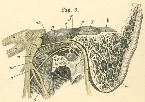
Hemifacial spasm (HFS) is mechanistically separate from BEB. The problem is not in the brain's motor signaling network - it is at the facial nerve root. A blood vessel presses against the facial nerve (CN VII) at the point where it exits the brainstem, generating constant irritation that causes the nerve to fire abnormally. The posterior inferior cerebellar artery and the anterior inferior cerebellar artery together account for the offending vessel in roughly three-quarters of cases.
Because the facial nerve supplies only one side of the face, HFS is always strictly unilateral - from first symptom to final presentation. It begins around the eye and spreads gradually downward over months to years: first the cheek, then the corner of the mouth, eventually the platysma in the neck. That slow downward progression is a recognizable clinical pattern.
The single most important feature distinguishing HFS from BEB is that hemifacial spasm persists during sleep. A bed partner who reports observing the movements overnight is providing the key piece of history. It reorients the entire evaluation away from a central dystonia and toward a peripheral nerve compression - and it points directly toward imaging of the posterior fossa as the next step.
Meige Syndrome: When Dystonia Spreads Beyond the Eyelids
Some patients with BEB develop involuntary movements of the jaw, tongue, or lower face - jaw clenching, tongue protrusion, lip pursing - alongside their eyelid spasms. This combination is Meige syndrome: BEB plus oromandibular dystonia. It represents a broader cranial dystonia, not simply a more severe version of eyelid spasm, and it requires a coordinated treatment approach across multiple facial territories. Patients whose symptoms extend below the eye level need a provider who can adjust injection maps to address the full distribution rather than limiting treatment to the periorbital muscles.
The Diagnostic Workup: Telling These Conditions Apart
No laboratory test confirms BEB, HFS, or apraxia of eyelid opening. The diagnosis is built from history, observation, and targeted testing in sequence:
- History of onset and pattern. When symptoms started, whether the problem is bilateral or strictly one-sided, whether spasms occur during sleep, and what reliably makes them worse or better.
- In-office observation under provocation. Bright light and sustained gaze can precipitate BEB spasms in the examination room. Sustained lateral gaze often provokes HFS. The clinician watches which muscles are active and whether the pattern is synchronous or clonic.
- Voluntary eye-opening test. The patient is asked to open their eyes on command. Failure to open in the absence of visible orbicularis squeezing - the eyelid sits at mid-position while the patient strains - suggests apraxia of eyelid opening rather than spasm driving the closure.
- EMG (electromyography). When clinical findings are ambiguous, or when prior Botox treatment has not worked as expected, surface or fine-wire EMG of the orbicularis and levator muscles distinguishes active spasm from levator inhibition. This is the definitive test for apraxia of eyelid opening.
- MRI with posterior fossa sequences. Ordered when HFS is suspected to identify the compressing vessel at the CN VII root exit zone. This imaging is standard before any referral for microvascular decompression and is not routinely needed for BEB.
Three patterns help orient the workup before testing begins:
- BEB: bilateral, stops during sleep, forceful orbicularis contraction visible during spasms
- HFS: strictly one-sided, persists during sleep, spreads progressively from eye toward lower face over time
- ALO: eyelid fails to open on command with no visible squeezing, often co-exists with BEB and explains Botox treatment failure
Apraxia of Eyelid Opening: the Condition Most Often Missed
Apraxia of eyelid opening (ALO) is a distinct and frequently overlooked condition in which the levator palpebrae superioris - the muscle that raises the upper lid - becomes inhibited, preventing voluntary eye opening even without active orbicularis contraction. The eyelid simply does not lift. There is no squeezing, no spasm. The patient strains to open and the eye stays shut.
ALO co-exists with BEB in a subset of patients and is confirmed by EMG demonstrating levator inhibition rather than orbicularis overactivity. Once confirmed, the injection strategy shifts: the goal becomes selectively reducing orbicularis tone in positions that allow the partially inhibited levator to work more effectively, rather than applying a standard spasm-reduction pattern across the periorbital muscles.
Botulinum Toxin Treatment: Injection Targets, Cycles, and Resistance
How the injections work and where they go
Botulinum toxin is the established first-line treatment for both BEB and HFS. Both indications are FDA-approved, and injections are typically covered by insurance when the diagnosis is properly documented with objective findings and functional impairment. Each cycle provides three to four months of relief, placing most patients on a schedule of three to four visits per year.
The injection maps differ between the two conditions - a detail that almost no practice-site content addresses. For BEB, the primary targets are three muscle groups: the orbicularis oculi, the corrugator supercilii, and the procerus. For HFS, modified patterns shift injection points away from the eyelid margin, using adjusted pretarsal and preseptal orbicularis positions along with modified brow-area placements. This shift reduces the risk of ptosis and dry-eye side effects that occur when toxin diffuses toward the levator or disrupts the lacrimal reflex - without reducing efficacy. Getting this right for HFS requires specific familiarity with toxin diffusion across different periorbital tissue planes.
Secondary resistance and formulation switching
A subset of patients who have responded well for years develop secondary resistance: injections wear off faster, and the symptom-free window shortens progressively. This is associated with an immune response to complexing proteins present in some toxin formulations - a low-grade neutralizing response that develops over repeated exposures. Switching to a different serotype or formulation can restore the therapeutic response. A formulation containing no complexing proteins, for instance, is thought to carry lower immunogenicity risk. Patients who notice their injection interval shortening should raise this with their provider rather than simply accepting more frequent dosing or assuming the condition has permanently worsened.
Surgical Options: Myectomy for BEB and Microvascular Decompression for HFS
| Feature | Orbicularis Myectomy (BEB) | Microvascular Decompression (HFS) |
|---|---|---|
| Target | Orbicularis oculi and adjacent muscles | Offending artery at CN VII root exit zone |
| Mechanism | Reduces muscle mass available for spasm | Removes compression - potentially curative |
| Ideal candidate | BEB inadequately controlled by Botox | HFS confirmed on MRI with significant functional impact |
| Published outcomes | Patient satisfaction above 90% | Roughly 90% spasm-free in large published series |
| Ongoing injections after surgery | Often eliminated; small lower-lid touch-ups remain in some cases | Generally not needed after successful procedure |
Orbicularis myectomy removes a portion of the orbicularis oculi and related periorbital muscles, reducing the contractile tissue available during a spasm. It is offered to BEB patients who have not achieved adequate control with botulinum toxin - whether from insufficient response, secondary resistance, or intolerance of injection frequency. Patient satisfaction in published outcomes exceeds 90%, and many patients can stop ongoing injections entirely. Full functional recovery takes up to six months, which means early post-operative results do not capture the final outcome.
Microvascular decompression (MVD) is the only treatment for HFS that addresses the cause rather than managing symptoms. A neurosurgeon accesses the posterior fossa through a small incision behind the ear, identifies the compressing vessel, and places a small Teflon sponge between the artery and the CN VII root to relieve the pressure permanently. In a published series of more than 5,000 cases, roughly 90% of patients were spasm-free at follow-up. MVD carries real neurosurgical risks - possible hearing changes, facial weakness, and standard posterior fossa surgical risks - and the decision requires a direct conversation between the patient, an oculoplastic surgeon, and a neurosurgeon. For patients with confirmed vascular compression on imaging and functionally limiting symptoms, MVD is the only intervention that can permanently resolve the condition rather than managing it cycle to cycle.
The Oculoplastic Surgeon's Role
Oculoplastic surgeons train in both ophthalmology and periorbital reconstructive surgery, building direct familiarity with levator and orbicularis function, lacrimal anatomy, and orbital mechanics. That anatomical base matters when injecting botulinum toxin near structures - the levator, the cornea, the lacrimal gland - where unintended diffusion causes ptosis, diplopia, or corneal exposure. It matters equally when evaluating whether a patient's problem is spasm, levator inhibition, a structural issue, or some combination.
For these eyelid movement disorders, the specialty manages the full arc of care:
- Confirming the diagnosis and distinguishing BEB from HFS from ALO from structural causes
- Initiating and adjusting botulinum toxin therapy, including recognition of secondary resistance and formulation changes
- Ordering and interpreting EMG when the clinical picture is ambiguous or treatment has underperformed
- Treating Meige syndrome with multi-territory injection maps that extend beyond the periorbital region
- Performing orbicularis myectomy for BEB patients who need surgical management
- Counseling HFS patients about MVD and facilitating the neurosurgical referral when imaging and symptom burden support it
Three conditions that close the eyelid against your will have three different causes. Treating the wrong mechanism solves nothing - and the workup that separates them is what makes the treatment work.
Frequently Asked Questions
How can I tell at home whether I have BEB or hemifacial spasm?
The clearest starting observations are laterality and sleep behavior. BEB affects both eyes and stops completely during sleep; hemifacial spasm affects only one side of the face and continues through the night. If a bed partner has noticed the movements while you sleep, that history points strongly toward HFS. These observations guide the evaluation but do not replace it - an in-person examination is required to confirm either diagnosis.
Why did my Botox injections stop working after years of good results?
Secondary resistance is the most likely explanation. Repeated exposure to complexing proteins in some botulinum toxin formulations can trigger a low-grade immune response that gradually neutralizes the drug. Switching to a different formulation - particularly one without complexing proteins - often restores the therapeutic response. If switching does not resolve the problem, apraxia of eyelid opening should be evaluated by EMG before drawing other conclusions about why treatment seems to be failing.
Is surgery only for severe cases?
For BEB, orbicularis myectomy is appropriate for patients not achieving adequate symptom control through injections - not strictly the most advanced cases only. Outcomes are strong, and many patients eliminate ongoing injection visits after surgery. For HFS, microvascular decompression is worth discussing relatively early if imaging confirms vascular compression and symptoms are affecting daily function, because it is the only option that addresses the underlying cause rather than managing symptoms on a recurring cycle.
Can dry eye cause blepharospasm?
Dry eye does not cause BEB, but it is a documented trigger that worsens existing spasms in a meaningful portion of patients. Treating underlying dry eye - with lubricating drops, punctal plugs, or prescription dry-eye therapy as appropriate - can reduce episode frequency and severity even before any change to the botulinum toxin regimen. Many patients benefit from managing both conditions at the same time rather than treating them as unrelated.
Will BEB or hemifacial spasm get worse if left alone?
BEB has a recognized natural history: it starts with mild increased blinking and progresses toward forceful, prolonged closures that prevent reading, driving, and basic navigation. In the most advanced stage, patients cannot keep their eyes open long enough for ordinary daily tasks despite having completely normal visual acuity. HFS spreads gradually from the periorbital region to the lower face and neck over months to years. Neither condition reverses on its own, which is the practical reason early evaluation and treatment planning matters.
What should I bring to a first appointment with an oculoplastic surgeon?
Come prepared with a written account of when symptoms began, whether the problem is one side or both, whether spasms continue during sleep or have been observed by a bed partner, and what makes episodes worse on a bad day. List every prior treatment - eye drops, injections, medications - and how your symptoms responded. The most useful thing you can describe is what the spasms prevent you from doing: the functional impact typically guides treatment decisions more directly than a description of the movements themselves.
General information only, not medical advice. Individual anatomy and healing vary. See the disclaimer.