The moment your blepharoplasty surgeon stretches your lower eyelid away from your eye, watches it drift slowly back into position, or stares at the inner corner of your lid while pressing gently outward, they are running a diagnostic test with direct consequences for your surgery. These maneuvers take perhaps 90 seconds total, but they determine whether your operation will include extra lid-tightening steps. Skip the testing, or fail to act on the results, and the most common serious complication of lower blepharoplasty follows: lid malposition, where the lower lid pulls away from the eye, exposes the white of the sclera, and leaves the cornea unprotected. The three core laxity tests - the snap-back test, the lateral distraction test, and the medial canthal tendon test - each use a grading system that maps directly to a surgical decision. Understanding them gives you a way to verify that your surgeon has assessed your specific anatomy, not just your appearance.
The Three Structures That Hold the Lower Eyelid Against the Eye
The lower eyelid does not stay in contact with the eyeball by accident. Three structures form its horizontal support framework, and all three loosen with age.
- The lateral canthal tendon anchors the outer corner of the lid to a small bony projection on the inner surface of the lateral orbital wall - approximately 4 to 5 mm behind the orbital rim - called Whitnall's tubercle. This tendon carries the greatest share of horizontal tension and is the first to stretch over time. As it attenuates, the lateral canthus drifts medially, the lid grows longer horizontally, and its contact with the globe loosens.
- The medial canthal tendon anchors the inner corner to the bone near the tear drainage opening. It opposes the outward pull of the lateral tendon and keeps the punctum - the small pore that drains tears - pressed against the eye. It fails independently of the lateral tendon and requires its own test.
- The tarsoligamentous sling is a sheet of fibrous tissue running the full horizontal length of the lid, tying the two canthal tendons together and providing the lid with its overall rigidity.
Involutional ectropion - the clinical endpoint of this process, where the lid rolls outward - is the most common form of ectropion seen in practice, precisely because this anatomical deterioration is universal. Descent of the malar fat pad with age withdraws mechanical support from below the lid, accelerating the progression. A surgeon examining a patient before blepharoplasty is trying to place that patient on this continuum before any incision is made.
The Snap-Back Test: Speed of Return as a Laxity Grade
How the test is performed
The surgeon grasps the central lower lid, displaces it gently downward away from the eyeball, then releases it. The patient is instructed not to blink. What the surgeon watches is how quickly - and whether - the lid returns to contact with the globe under its own passive elasticity.
What each grade means
The snap-back test uses a five-point grading scale from 0 to IV. Grade 0: the lid returns to the globe immediately on release - normal elasticity, no concern. Grade I: return takes 2 to 3 seconds - mild laxity, unlikely to cause problems on its own. Grade II: return takes 4 to 5 seconds - moderate laxity, warrants careful consideration especially when other risk factors are present. Grade III: the lid takes more than 5 seconds to return, but does return after the patient blinks - significant laxity that frequently requires preventive canthal support. Grade IV: the lid never returns to the globe even after blinking - this constitutes frank ectropion already present before surgery, and canthal reconstruction is mandatory.
The blink instruction and why it matters
Asking the patient not to blink is not a formality. Blinking activates the orbicularis muscle, which actively pulls the lid back to the globe and masks the true passive elasticity being tested. A Grade III lid can easily register as Grade I if the patient reflexively blinks the moment the surgeon lets go. Examiners typically give a clear instruction before releasing: keep your eyes open, do not blink until told. Any result obtained without this instruction is unreliable.
The Lateral Distraction Test: Measuring Laxity in Millimeters
Where the snap-back test measures the speed of return, the lateral distraction test measures distance. The surgeon grasps the central lower lid and draws it anteriorly - away from the eyeball - noting the displacement in millimeters before meaningful resistance is felt. The four ranges correspond to increasing surgical urgency: 0 to 2 mm is normal; 2 to 4 mm indicates mild laxity; 4 to 6 mm indicates moderate laxity; greater than 6 mm indicates severe laxity. When displacement exceeds 8 mm and is combined with a significantly delayed snap-back, most surgeons treat intraoperative canthal support as non-negotiable.
The traditional manual pull test has long been subject to inter-examiner variability - different hand sizes and different applied forces can shift measurements. A 2021 study introduced a three-dimensional stereophotogrammetry approach specifically to address this problem, and found that structured, objective measurement produced far more consistent results between examiners than unguided manual pull. The direction the field is moving is toward reproducible numbers rather than subjective clinical feel.
| Laxity Level | Snap-Back Grade | Distraction Distance | Typical Surgical Response |
|---|---|---|---|
| None | Grade 0 | 0-2 mm | No canthal support added |
| Mild | Grade I | 2-4 mm | Monitor; canthopexy if other risk factors present |
| Moderate | Grade II | 4-6 mm | Preventive canthopexy often warranted |
| Significant | Grade III | Greater than 6 mm | Canthopexy or tarsal strip canthoplasty required |
| Frank ectropion | Grade IV | Greater than 8 mm (severe) | Formal canthoplasty mandatory |
The Medial Canthal Tendon Test: The Second Anchor
This test receives far less attention in patient-facing resources than the snap-back or distraction test, which is a genuine gap - because the medial canthal tendon fails independently, and lateral tightening alone cannot fix a loose inner corner.
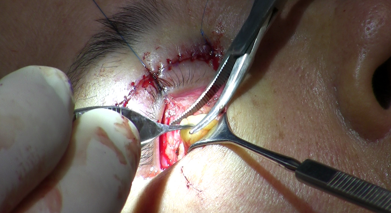
The surgeon grasps the lower lid and displaces it toward the outer corner of the eye, watching how far the punctum - the small drainage opening at the inner corner - moves from its resting position. In a normal lid, punctal movement is no more than 1 mm. A displacement of 2 mm reflects mild laxity of the medial canthal tendon; greater displacement indicates progressively more laxity that may require direct surgical attention. Confirm the exact threshold your surgeon uses, as grading conventions vary.
The clinical implication is specific. A lateral tarsal strip procedure, which tightens the outer anchor, will not address a loose inner one. Applying tension only to the lateral end of a lid with bilateral laxity can produce an abnormal contour - tight at the outer corner, still loose in the center - and ongoing tear drainage problems as the punctum continues to drift away from the eye. When medial laxity is confirmed, the surgical plan must expand to include a medial canthoplasty or plication suture targeting the medial tendon directly.
What Else the Surgeon Records at the Same Visit
The three canthal tests do not operate in isolation. Several complementary measurements are gathered at the same consultation and combined with laxity grades to build a complete preoperative risk picture.
- MRD2 (marginal reflex distance 2): the distance from the corneal light reflex - the white dot produced by a penlight - to the lower lid margin in straight-ahead gaze. The commonly cited normal value is approximately 5 to 5.5 mm. A value above roughly 5.5 mm suggests the lower lid may already be sitting lower than expected before any surgery, indicating baseline retraction or lid-globe separation; confirm the exact threshold with your surgeon as reference ranges vary slightly by source. This becomes the documented starting point against which postoperative outcomes are measured.
- Scleral show: visible white between the iris and the lower lid margin in primary gaze. Some patients have mild baseline scleral show from prominent eyes or a naturally low lid position, but in the blepharoplasty evaluation context, documented scleral show is an independent risk factor for postoperative lid malposition after transcutaneous surgery - it is recorded separately from laxity, not as a substitute for testing it.
- Orbital vector: assessed by viewing the patient in profile and noting whether the cornea projects forward of the inferior orbital rim. A negative orbital vector - cornea anterior to the rim - means the lower lid lacks bony support below it. Any downward pull on the lid after surgery meets less skeletal resistance. When a negative orbital vector and moderate laxity are both present, the two risk factors compound each other, and the surgical plan reflects both.
How Each Finding Translates Into the Operating Room
The three surgical pathways
Grades 0 and I with no additional risk factors: no canthal support is added. The blepharoplasty proceeds with fat and skin management as planned, and the lid's existing elasticity is sufficient to maintain position.
Grade II, or borderline findings combined with scleral show, negative orbital vector, or a planned transcutaneous incision: a preventive canthopexy is performed. A suture is passed through the lateral canthal tendon or the periorbita at the orbital tubercle to resuspend the canthus without fully detaching it. This adds minimal time to the operation and addresses a predictable problem before it occurs.
Grades III and IV: a lateral tarsal strip canthoplasty is required. The lateral canthal tendon is split at the outer corner, epithelium is stripped from a short segment of the posterior lid margin, and the exposed strip of tarsus is anchored directly to periosteum at the lateral orbital tubercle. This physically shortens and repositions the lower lid - a structural reconstruction for structural failure.
Why the incision approach shifts the threshold
The transconjunctival approach leaves no external scar and therefore carries substantially lower malposition risk. It cannot remove excess skin, which is why transcutaneous surgery is sometimes necessary - but that benefit comes with a measurable increase in risk. Surgeons working through a skin incision raise their index of suspicion for canthal support across every laxity grade because the downstream force on the lid is greater.
The decision sequence a surgeon works through before finalizing the plan:
- Record snap-back grade and lateral distraction measurement.
- Perform the medial canthal test and note any medial displacement.
- Measure MRD2 and document any baseline scleral show in primary gaze.
- Assess orbital vector by profile view.
- Confirm whether the planned incision is transconjunctival or transcutaneous.
- Combine all five findings to determine the support requirement: no additional support, preventive canthopexy, or formal canthoplasty.
What Happens When Laxity Is Missed
The single most common cause of postoperative lower lid malposition after lower blepharoplasty is failure to identify and address preoperative laxity. The complication does not appear all at once - it progresses through a sequence that begins with a mild cosmetic change and ends with a medical problem.
- The lower lid sits lower than expected in the days to weeks after surgery - MRD2 increases, more scleral white is visible.
- The lid stops touching the globe consistently. Tear drainage is disrupted. Dry-eye symptoms, tearing, and chronic irritation develop.
- As the lid continues to retract or rotate outward, frank ectropion establishes - the conjunctiva is chronically exposed, and the cornea is at risk for breakdown.
Revision surgery for this complication is considerably harder than prevention. Scar tissue in both the anterior lamella (skin and muscle) and posterior lamella (conjunctiva and tarsus) limits access and tissue mobility. A tarsal strip that would have been straightforward to place during the original operation now requires dissection through contracted planes. Skin grafts or spacer grafts are sometimes necessary to restore vertical lid height when scarring has shortened the anterior lamella. The revision outcome, even in expert hands, rarely matches what a correctly planned primary surgery could have achieved.
The time to address lid laxity is at the primary surgery. Correcting postoperative malposition means operating through scar with fewer tissue options and a narrower margin for error - prevention is not a supplementary precaution, it is the standard of care.
Questions to Ask at Your Lower Blepharoplasty Consultation
Most consultation pages promise a thorough evaluation without specifying what that evaluation contains. These questions give you a concrete way to confirm that laxity testing was performed, graded, and factored into your surgical plan - not just observed vaguely.
- Did you perform a snap-back test, and what grade did my lids score? What does that grade mean for my plan?
- How far could you pull my lower lid away from the eye - where does that measurement fall in the normal range?
- Did you test the medial corner of my lid as well, or only the outer corner?
- What is my MRD2, and is my lower lid already sitting lower than ideal before surgery?
- Do I have a negative orbital vector, and how does that combine with my laxity findings?
- Are you planning to add a canthopexy or canthoplasty to my surgery, and which technique? Why or why not?
- If you are using a skin incision, how does that change your threshold for canthal support in my specific case?
A surgeon who has performed this testing will answer each of these questions specifically, citing the grades and measurements they recorded. A vague answer - "your eyes look fine" or "we'll assess things in the operating room" - is worth following up on with a direct question about which tests were done and what they showed.
Frequently Asked Questions
Can I detect lower eyelid laxity myself before seeing a surgeon?
Most patients cannot grade their own laxity at home. You might notice that more white is visible below the iris than it used to be, or that a lower lid looks slightly lower than its partner, but these findings are subtle until laxity is advanced. The critical information - how the lid behaves under displacement - requires hands-on testing. Looking in a mirror only shows the lid at rest.
If my snap-back grade is II, does that automatically mean I need a canthoplasty?
Grade II typically points toward preventive canthopexy rather than formal canthoplasty, but the decision also depends on your distraction measurement, MRD2, orbital vector, and planned incision route. The same Grade II result in a transcutaneous case may warrant canthopexy, while the same grade in a transconjunctival patient with no other risk factors might be observed. Ask your surgeon how the findings combine in your specific situation, not just what one test showed.
Will adding canthal support change how my eyes look after surgery?
A correctly performed canthopexy or lateral tarsal strip is intended to hold the lid where it anatomically belongs, not to produce a visibly altered appearance. The goal is support, not repositioning above the natural level. In patients whose lateral canthus has already drifted medially from laxity, a tarsal strip can actually improve the outer canthal angle slightly - restoring, not creating, a normal contour.
What is the practical difference between a canthopexy and a canthoplasty?
A canthopexy resuspends the lateral canthal tendon without dividing it - a suture repositions the structure while it remains intact. A canthoplasty divides the tendon, modifies the tissue, and reattaches it; the lateral tarsal strip technique is the standard approach for higher-grade laxity. Canthopexy suits prevention in borderline cases; canthoplasty is used when the laxity is significant enough to require structural reconstruction rather than repositioning alone.
Does the surgeon recheck laxity in the operating room, or only before surgery?
Most surgeons reassess the lid under anesthesia before making incisions, because muscle relaxation and patient positioning both affect what the lid does. A Grade II lid in the office can appear more lax on the table. The preoperative grading sets the expectation and informs consent; the intraoperative check confirms whether the plan needs to be adjusted before the first incision.
Is there any reason a surgeon might not perform laxity testing?
In an experienced oculoplastic or facial plastic surgery practice, laxity testing before lower blepharoplasty is standard. It might be omitted in very brief consultations, in practices where lower lid surgery is occasional rather than routine, or if the consultation focuses on cosmetic concerns without examining lid function. The tests take under two minutes; if they were not mentioned, asking directly whether they were performed is a reasonable step before finalizing a surgical plan.
General information only, not medical advice. Individual anatomy and healing vary. See the disclaimer.