When someone comes in for upper eyelid surgery and mentions puffiness at the outer corner, the assumption - from the patient, sometimes from other providers - is excess fat or loose skin. Often that is correct. But roughly one in ten people presenting for upper blepharoplasty has a different problem entirely: the lacrimal gland, a small secretory organ that normally sits at the roof of the outer orbit, has slipped forward into the visible portion of the upper lid. The bulge it creates looks superficially like fat herniation. It is not, and treating it as though it were fat is one of the more consequential mistakes in cosmetic eyelid surgery.
What the Lacrimal Gland Is and What It Does
The lacrimal gland is the primary source of the watery layer of your tear film - the aqueous component that makes up the bulk of every tear. Without it, the eye surface dries out rapidly, becomes inflamed, and eventually sustains damage. The gland is not passive; it secretes continuously throughout the day and responds to reflex stimulation such as dust, bright light, and emotion.
Anatomically, the gland has two lobes. The larger one - the orbital lobe - sits inside the orbit in a shallow bony depression called the lacrimal fossa, located at the outer roof of the orbit just inside the rim of the frontal bone. The smaller palpebral lobe, significantly smaller than the orbital lobe, lies beneath the levator aponeurosis in the lateral upper eyelid itself; a surgeon can see it directly by everting the upper lid and looking at the outer segment of the conjunctival surface.
Both lobes drain through excretory ducts - six to twelve from the orbital lobe and two to five from the palpebral lobe - all emptying into the superior conjunctival fornix, the pocket where the upper eyelid lining meets the eyeball. This drainage anatomy becomes critically important when the question of removal arises.
Why the Gland Moves Forward
Two supporting structures normally hold the orbital lobe in the lacrimal fossa. Fibrous bands called Soemmerring's ligaments run from the gland capsule to the periosteum, anchoring it to the orbital roof. The primary support, though, is Whitnall's ligament - a condensation of orbital fascia that functions as a hammock, suspending the orbital lobe from above. When Whitnall's ligament is intact and taut, the gland stays recessed. When it stretches or develops small tears over years, the gland drifts forward, pressing against the orbital septum and eventually bulging into the lateral upper lid space.
This is predominantly an age-related process. Ligamentous laxity throughout the periorbital region is a consistent finding as patients age, and Whitnall's ligament is not exempt. A 2025 study published in BMC Ophthalmology confirmed that gland size and anterior gland position both correlate with how visible prolapse becomes - meaning patients with naturally larger glands are more likely to develop noticeable displacement as support weakens.
Prolapse also appears in specific clinical settings beyond normal aging: Floppy Eyelid Syndrome, thyroid eye disease, and chronic orbital inflammatory conditions all stress these supporting structures in distinct ways. When any of those underlying diagnoses is present, the surgeon needs to identify and address it in the correct sequence. Operating on the gland without managing active thyroid eye disease, for example, undermines both the safety and durability of any repair.
What the Patient Sees and Feels
The most consistent complaint is a soft fullness at the outer third of the upper eyelid - not the middle, not the inner corner, but specifically the temporal zone. Patients describe it as a lump, a heaviness, or a bulge that does not match the drooping they notice elsewhere in the lid. Some have been told it is simply excess fat and to expect improvement after standard blepharoplasty. When surgery proceeds without addressing the gland, the lateral fullness remains and the patient is left wondering why the result is incomplete.
The S-Shaped Contour
Looking straight ahead in good light, a patient with lacrimal gland prolapse often shows what surgeons call an S-shaped upper lid contour. The central lid follows a smooth, concave arc inward - the levator muscle is giving proper support there - but the outer lid bows convex where the gland is pushing forward. The silhouette traces a gentle S rather than a clean, continuous arc. This sign is not produced by skin excess alone and not by fat herniation alone, because the lateral upper lid has no fat compartment at the location where the gland sits. Focal convexity limited to the outer third is the key visual clue.
Signs That Point Toward Gland Rather Than Fat
- Fullness confined to the outer third of the upper lid, with the central and nasal lid appearing relatively flat
- A bumpy or lobulated texture when the outer lid is pressed gently through closed skin
- Swelling that is noticeably worse after crying or after rubbing the eye
- Greater prominence in the evening compared with the morning
- A bulge that persists or even increases when looking straight ahead with the brow elevated
Unlike skin excess, which is constant and static, gland prolapse fluctuates. The gland swells after lacrimal stimulation, after prolonged screen use, and after eye rubbing. That variability is itself a diagnostic clue that the structure involved is secretory tissue, not inert herniated fat.

How Common This Finding Actually Is
Clinical detection at consultation identifies lacrimal gland prolapse in roughly 10 to 15 percent of people presenting for upper blepharoplasty. That figure understates the true prevalence. When surgeons open the upper lid and examine the anatomy directly, prolapse rates in older patients reach 60 percent. The discrepancy exists because mild and moderate displacement does not always produce obvious surface changes - it takes operative exposure to see exactly where the gland sits relative to the orbital rim.
One published series of 198 consecutive upper blepharoplasty patients found 20 cases - 10.1 percent - with clinically significant prolapse, and 70 percent of those patients were asymptomatic preoperatively. The severity distribution across mild, moderate, and severe categories is reported in the primary publication (Lacrimal gland prolapse in upper blepharoplasty, Orbit, 2019); verify those proportions there before citing them. A surgeon who only investigates when the patient specifically raises outer fullness will miss a substantial number of cases that are present and visible intraoperatively. Thorough evaluation means examining the lateral lid in every upper blepharoplasty candidate.
How a Surgeon Makes the Diagnosis
Diagnosing lacrimal gland prolapse combines inspection, palpation, a positional test, and in some cases eyelid eversion. No imaging is required in clear presentations, but CT or MRI is ordered if the findings are atypical or if the surgeon needs to rule out an orbital mass.
Inspection and Palpation
The surgeon looks at the upper lid contour in primary gaze, specifically for a lateral convexity or S-shaped silhouette. Gentle pressure at the outer upper lid through closed skin then assesses what is sitting there. Orbital fat is soft, smooth, and compressible - it yields readily. The lacrimal gland is different: lobulated on its surface, firmer, and does not compress as freely. In many cases, light sustained pressure actually reduces the gland back into the lacrimal fossa and the bulge temporarily disappears - a finding that cannot be replicated with herniated fat.
The Supine Test
This is the most useful bedside maneuver for distinguishing gland prolapse from fat prolapse. The patient lies flat, looks downward, and the brow is gently elevated to relax the orbicularis. In that position, herniated orbital fat redistributes into the orbit and the bulge decreases or disappears. The lacrimal gland does not redistribute, because it is a solid organ with fibrous attachments, not free fat. Any lateral fullness that persists when the patient is supine is presumed gland until proven otherwise.
Eyelid Eversion
The upper lid is gently everted so the conjunctival surface faces outward. At the outer edge of the everted lid, the palpebral lobe appears as a pinkish-gray, slightly lobulated structure against the pale pink conjunctiva. This view gives the surgeon direct information about lobe size and position that the external surface cannot provide.
| Feature | Lacrimal Gland Prolapse | Orbital Fat Herniation | Skin and Tissue Excess |
|---|---|---|---|
| Location in upper lid | Outer third only | Central and inner thirds | Full lid width |
| Texture on palpation | Lobulated, firmer | Soft, smooth, compressible | Movable skin folds |
| Supine test result | Persists lying flat | Reduces lying flat | Unchanged by position |
| Color when visible | Pinkish-gray, lobulated | Yellow | Normal skin tone |
| Lid contour effect | S-shaped curve laterally | General fullness, no S-curve | Diffuse droop, no focal convexity |
What a Missed Diagnosis Costs
Beyond cosmetic disappointment, the more serious risk is accidental excision. A prolapsed lacrimal gland sitting in the lateral compartment can look, at a glance, like an aberrant fat pad to a surgeon who is not actively looking for it - especially if the preoperative examination did not identify it. Dissection through the orbital septum in that zone can encounter gland tissue before the surgeon registers what it is. If that tissue is removed, the patient loses secretory function that cannot be restored.
Why the Gland Can Never Be Removed
The lacrimal gland produces the aqueous layer of the tear film. Without that layer, the tear film breaks down within seconds after each blink. The result is aqueous-deficient dry eye syndrome: persistent burning, stinging, and gritty discomfort; sensitivity to air conditioning and screens; fluctuating blurry vision from an unstable tear surface; and, in untreated cases, corneal surface damage.
The anatomy of the drainage system makes even partial removal damaging. All of the ducts from the orbital lobe pass through the palpebral lobe on their way to the superior conjunctival fornix. Removing the palpebral lobe alone therefore disrupts secretion from the entire gland above it - not just the tissue that was excised. Published case reports in the ophthalmic literature have documented refractory aqueous-deficient dry eye following inadvertent excision of palpebral lobe tissue that was mistaken for another structure; consult your oculoplastic surgeon or the primary literature for specific cases. The patient's dry eye was permanent and unresponsive to standard treatment.
Every duct from the orbital lobe passes through the palpebral lobe before reaching the eye. Removing any portion of the gland without understanding this architecture risks eliminating tear secretion from the entire structure.
There is no reconstructive option for lost lacrimal gland tissue. Lubricating drops, punctal plugs, and prescription tear-stimulating medications can reduce symptoms but do not restore the secretion that was present before surgery. This is why identification before the first incision is not optional.
How Dacryoadenopexy Repairs the Problem
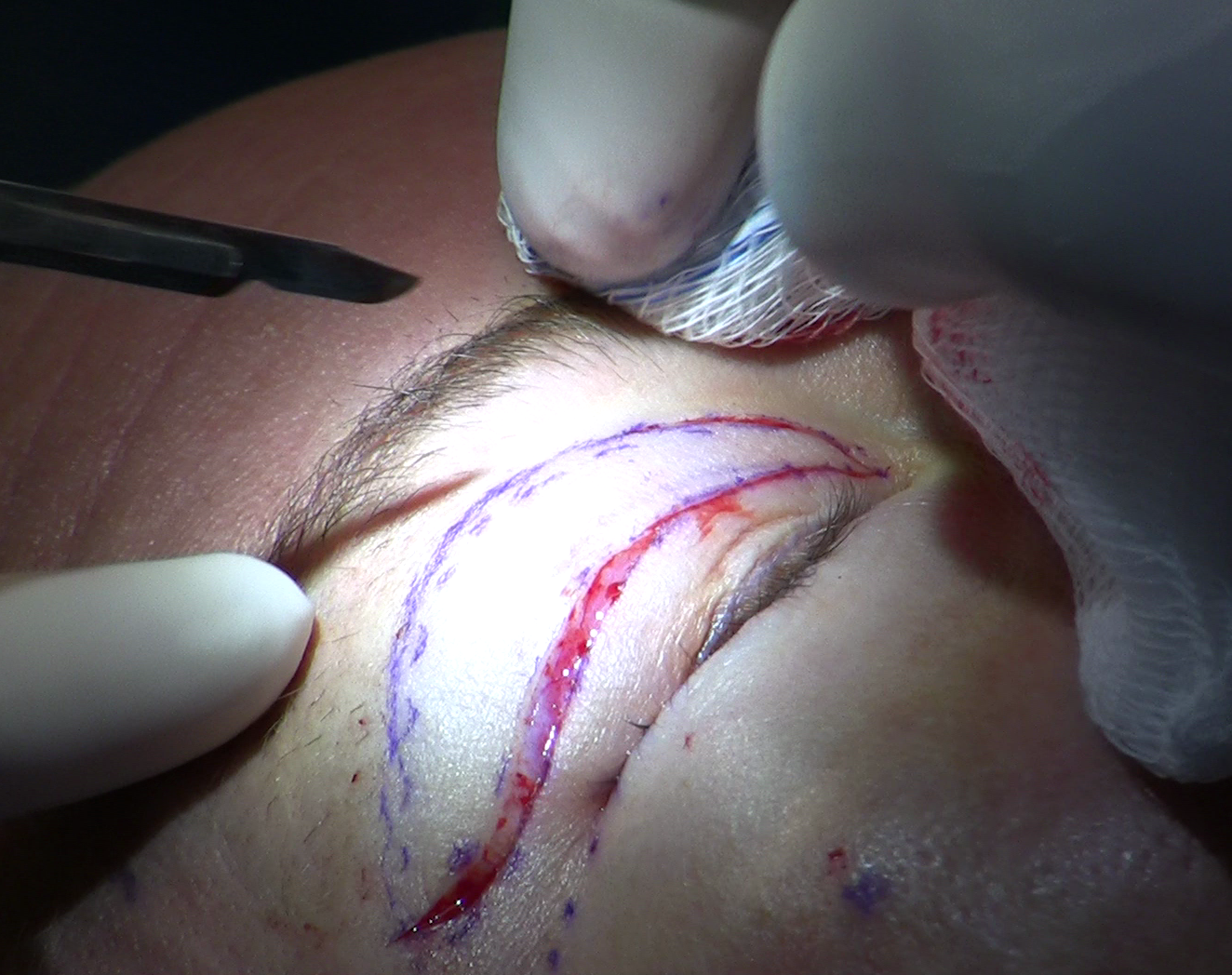
Dacryoadenopexy - surgical fixation of the lacrimal gland - restores the gland to the lacrimal fossa and anchors it there. It is performed through the same upper blepharoplasty incision used for the cosmetic portion of the procedure, so no additional external scar results. The two structures form a single coordinated operation.
- The surgeon marks and creates the standard upper blepharoplasty skin incision at the lid crease, extending laterally as needed toward the outer canthus.
- Dissection proceeds through the orbicularis muscle and orbital septum. The prolapsed orbital lobe is identified in the lateral compartment - pinkish-gray, lobulated, firmer than surrounding fat.
- The gland is gently freed from any adhesions that formed as it migrated forward, taking care to avoid the ductules running through the tissue.
- The orbital lobe is repositioned into the lacrimal fossa and manually confirmed to seat correctly against the periosteum of the frontal bone.
- Fixation is achieved with one of two main techniques: a double-armed nonabsorbable monofilament suture (nylon or Prolene, with gauge - commonly 4-0, 5-0, or 6-0 - selected by the surgeon based on tissue) passed through the inferior gland capsule and tied to the periosteum of the superior orbital rim, or a Whitnall's ligament sling in which the ligament itself is sutured over the anterior gland surface and fixed to the orbital rim periosteum - replicating the natural hammock mechanism that had failed.
- The blepharoplasty portion continues: skin and any excess orbicularis are removed, central fat compartments are managed, and the incision is closed in layers.
Both fixation approaches are described in the oculoplastic literature and, in published series, show low relapse rates; however, long-term comparative outcome data remain limited, so discuss technique choice and expected durability directly with your surgeon. Recovery mirrors standard upper lid blepharoplasty recovery - bruising and swelling for one to two weeks, with most patients presentable socially by ten to fourteen days. The outer upper lid, previously the source of persistent lateral fullness, flattens as swelling resolves and the repositioned gland settles. Tear function is preserved.
What to Ask at a Blepharoplasty Consultation
Evaluation for lacrimal gland prolapse should be a routine part of every upper blepharoplasty assessment, not something reserved for patients who specifically raise it. You can confirm it is being done by asking directly:
- Are you examining the outer upper lid specifically for lacrimal gland prolapse, not just excess skin and fat?
- Will you use the supine test to distinguish gland prolapse from orbital fat herniation?
- If prolapse is found, which fixation technique do you use - periosteal suture, Whitnall's ligament sling, or another approach?
- Do I have any findings - thyroid eye disease, Floppy Eyelid Syndrome - that should be evaluated before eyelid surgery is scheduled?
- If prolapse is identified intraoperatively but was not planned for preoperatively, how do you handle that decision during the procedure?
Frequently Asked Questions
Can lacrimal gland prolapse improve on its own without surgery?
No. The ligamentous laxity that allows the gland to migrate forward does not reverse without surgical repair. The prominence may vary day to day - swelling after crying, for example, or being less noticeable in the morning when you have been lying flat - but the underlying displacement is permanent. Conservative treatments such as lubricating drops or anti-inflammatory medications do not restore the gland to its normal position.
Will dacryoadenopexy affect my tear production or give me dry eye?
Repositioning surgery does not remove gland tissue, so it does not reduce tear secretion. Most patients experience the short-term surface dryness common after any eyelid surgery - from reduced blinking and minor swelling - which resolves within a few weeks. Patients who already have mild dry eye before surgery should disclose that at consultation, since it affects both the surgical plan and the expected postoperative course.
What if I already had blepharoplasty and the outer fullness is still there?
Persistent lateral upper lid fullness after blepharoplasty is one of the clearest postoperative presentations of a prolapse that was not identified preoperatively. Return to your surgeon or seek an oculoplastic second opinion. If the gland is intact and accessible, dacryoadenopexy can be performed as a revision procedure. The options and complexity depend on exactly what the original surgery did and did not address in that zone.
Is the prolapse usually in both eyes or just one?
Both eyes are commonly involved, even when only one side is visually prominent. Age-related laxity of Whitnall's ligament affects both orbits, though the degree of displacement can be asymmetric. A thorough evaluation covers both lids regardless of which side prompted the visit, since correcting one side while leaving the other untreated produces a mismatched result after surgery.
How does thyroid eye disease affect this condition?
Thyroid eye disease causes soft tissue inflammation and expansion within the orbit, which can push the lacrimal gland forward and disrupt its supporting attachments. When thyroid eye disease is active, operating on the gland produces unpredictable results because the orbital environment continues to change. A surgeon evaluating a blepharoplasty candidate with known thyroid disease - or with signs such as lid retraction or prominent eyes - will coordinate with an endocrinologist to confirm the disease is stable before scheduling any periorbital procedure.
Is a prolapsed lacrimal gland dangerous if left alone?
Left untreated, lacrimal gland prolapse does not threaten vision or tear function - the gland continues to secrete normally even when displaced forward. The risks are cosmetic (persistent outer lid fullness) and surgical (accidental excision if a future operation encounters the gland without recognizing it). Any patient who is a blepharoplasty candidate and has documented prolapse should have it addressed at the time of lid surgery rather than deferred to a later procedure.
General information only, not medical advice. Individual anatomy and healing vary. See the disclaimer.