Most patients walk away from blepharoplasty expecting a visible scar for months. Many are surprised when, within a few weeks, they struggle to find the incision line at all - even in a mirror. That outcome is not luck. It follows directly from where the cut is placed, what eyelid skin is made of, and how the lid physically moves when the eye opens. Understanding those mechanics, and knowing what the healing timeline actually looks like, takes a significant amount of the uncertainty out of recovery.
Why Eyelid Skin Heals Differently from Everywhere Else
Upper eyelid skin is approximately 0.5 mm thick - the thinnest skin anywhere on the human body. That single fact shapes everything that follows in healing. When the body repairs a wound, it fills the gap with collagen. On thick skin - a knee, a shoulder, the back - there is room for collagen to pile up into a raised, firm ridge. On the eyelid, there is almost no structural space for that to happen. The amount of collagen the body can deposit is physically limited by the tissue depth, which is why the final scar line on the eyelid is typically finer and flatter than virtually any other surgical scar.
The periorbital area is also richly vascularized. More blood supply means more oxygen and growth factors delivered to the wound bed, faster epithelialization of suture tracks, and quicker resolution of inflammation. The same vascularity that causes dramatic bruising in the first days after surgery is working in the patient's favor by day four.
One additional factor: the eyelid is a moving structure. Constant gentle movement along the incision line during normal blinking maintains pliability in the healing tissue and discourages the rigid cross-linking that produces contracture-style scarring in immobilized tissue. The combination of thin skin, rich blood supply, and continuous gentle movement makes the eyelid one of the most scar-forgiving surgical sites on the body.

Upper Lid Incision Placement and the Dynamic Mechanics of Concealment
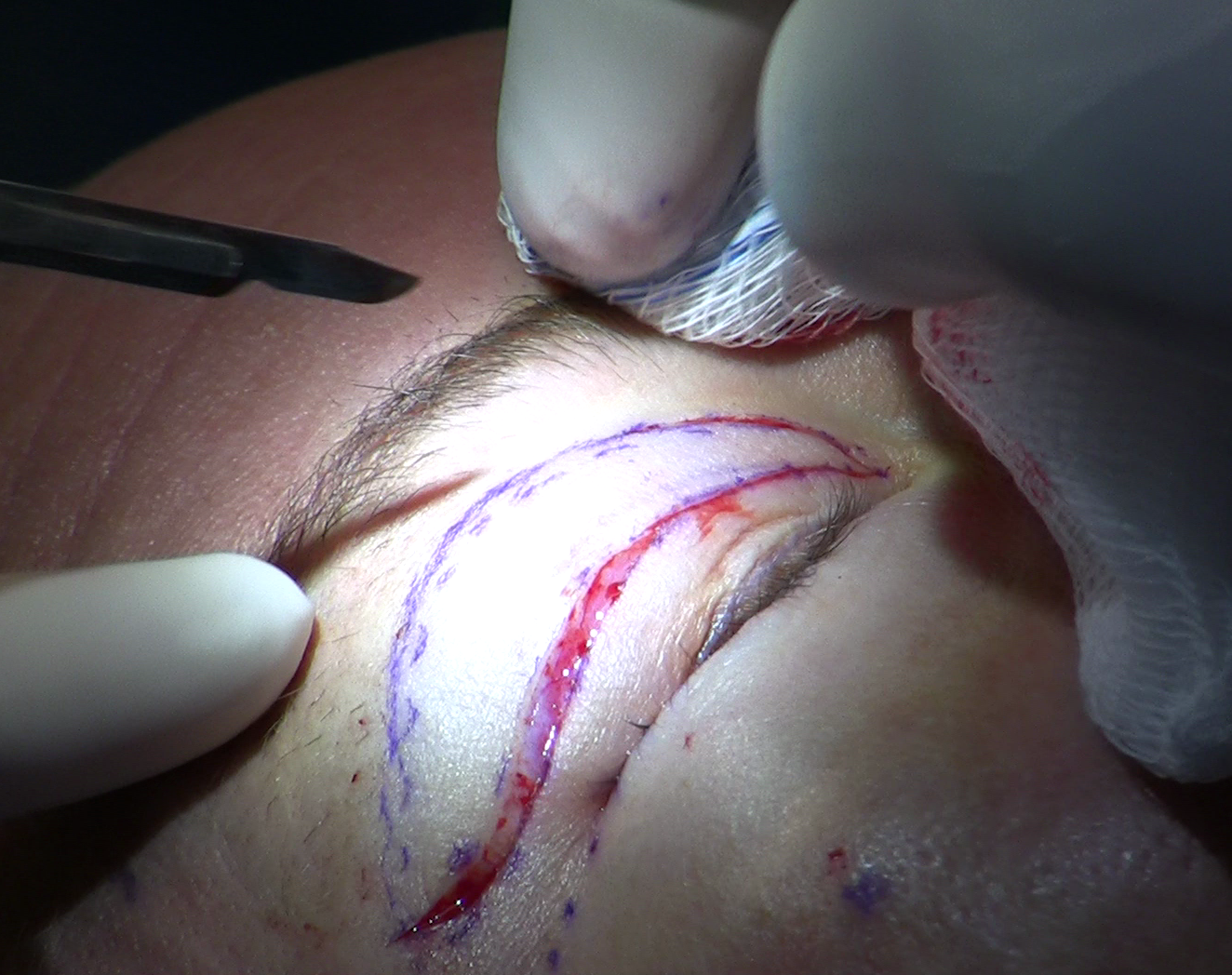
The upper blepharoplasty incision is placed in the supratarsal fold - the natural crease that forms where the levator aponeurosis, the muscle that lifts the lid, attaches to the overlying skin. In women this fold typically sits 7-10 mm above the lash line; in men the landmark is slightly lower, around 7-8 mm. The surgeon traces the existing natural crease rather than choosing an arbitrary line.
What competing explanations of this technique consistently miss is the dynamic anatomy. When the eye is closed or at rest, the crease is simply a fold in the skin. When the eye opens, the levator muscle contracts and the upper lid rises. As it rises, the skin above the crease folds over and drapes downward across the incision line. The fold does not merely sit near the scar - it actively moves over it each time the eye opens, functionally hiding the incision at conversational distance for the vast majority of patients. The eye-open position is how people see you in daily life. The concealment is structural and mechanical, not coincidental.
Lower Lid Incisions: Two Routes, Two Very Different Scar Profiles
Lower blepharoplasty offers a genuine choice between two approaches with fundamentally different scar profiles - not just slightly different ones.
| Feature | Transconjunctival | Subciliary |
|---|---|---|
| Incision location | Inner surface of lower lid | External skin, 1-2 mm below lash line |
| External scar | None | Present, heals at lash margin |
| Best suited for | Good skin tone, fat treatment only | Excess skin requiring excision |
| Post-inflammatory hyperpigmentation risk | None | Real risk in darker Fitzpatrick types |
| Patient selection demands | More forgiving | Requires precise anatomy assessment |
Transconjunctival lower blepharoplasty is not merely a variation in technique - it is a scar-elimination strategy. The incision is made entirely on the inner conjunctival surface of the lid, leaving no external wound and therefore no external scar, no suture tracks on the skin, and no hyperpigmentation risk at any incision site. For patients with adequate lower-lid skin tone who need fat repositioning or removal only, this approach removes scar risk from the equation entirely. It is worth framing it that way in the consultation.
The subciliary route is chosen when the lower lid has true excess skin that needs excision. Placed 1-2 mm below the lash line, the healed incision sits within the natural shadow of the lashes and, in patients with good tissue quality and consistent sun protection, typically becomes difficult to distinguish from the lash margin. It requires more careful patient selection and carries real considerations for patients with darker skin tones.
The Three Phases of Wound Healing on Eyelid Skin
Inflammatory Phase: Days 1 Through 3
The incision triggers an immediate vascular response - histamine release, capillary dilation, and white cell recruitment. Clinically this looks like swelling, redness, warmth, and bruising. This phase is working correctly when it looks alarming. On the eyelid, the rich vascularity that makes it look intense also helps it resolve faster than on less well-perfused tissue.
Proliferative Phase: Day 3 Through Week 6
Fibroblasts migrate into the wound and begin laying down collagen. New capillaries grow into the healing tissue - neovascularization - which is why the scar appears its pinkest and may feel slightly firm or raised during this window. This is the phase patients most often worry about. A pink, slightly thickened incision line at week three is normal proliferative biology, not a sign of abnormal scarring.
Remodeling Phase: Week 6 Through Month 12
Collagen fibers reorganize from a random, immature weave into a more ordered structure. The scar flattens progressively, the pink color fades to skin tone and eventually to pale, and the tissue softens. This phase cannot be rushed. Final scar quality should not be assessed - by the patient or the surgeon - until at least six months post-operatively. Many scars continue improving through the full 12-month mark.
What the Scar Looks Like Week by Week
Knowing the expected visual progression helps patients distinguish normal healing from genuine problems:
- Days 1-3: Swelling peaks, bruising spreads (often to the cheek), incision line is red and clearly visible.
- Days 5-7: Sutures are removed. The track holes are briefly open; this is the window when milia can begin forming.
- Weeks 2-3: Bruising resolves, swelling decreases substantially. The incision remains pink and may feel slightly firm.
- Weeks 4-6: The scar reaches its most visually prominent phase - still pink, still palpable. Normal peak proliferative activity. This is the stage that triggers the most unnecessary concern.
- Months 2-4: Pinkness begins fading. The crease looks increasingly natural. Most patients are comfortable in social settings by this point.
- Months 6-12: Ongoing maturation. Color approaches surrounding skin tone, scar flattens fully. The result at 12 months is the true final outcome.
Milia Are Not Scars
The most common complaint patients describe as a scar problem after blepharoplasty is milia - and milia are not scar tissue at all. They are tiny white cysts formed when keratin becomes trapped inside a healing suture track. Each suture needle creates a small entry and exit hole in the skin. During the brief window between suture removal at days 5-7 and full closure of those track holes, a flap of surface epidermis can fold inward and seal keratin inside the tunnel. The result is a small white or yellowish dome sitting precisely at the former suture mark.
Milia have none of the characteristics of a scar. They do not raise the tissue along the incision line, they do not contract, they do not darken with sun exposure. Many resolve spontaneously within weeks. Those that persist are handled in the office with a sterile needle - a quick pricking maneuver that evacuates the cyst contents, leaves no mark, and requires no anesthesia.
The practical distinction between milia and a developing hypertrophic scar:
- Milia are round, white, and dome-shaped, sitting at specific old suture points rather than along the full incision
- A hypertrophic scar is linear, pink or red, and follows the entire incision path as a raised ridge
- Milia do not feel different from surrounding skin when gently palpated
- Hypertrophic tissue feels firm and raised when pressed along the incision line
- Milia typically appear within 2-4 weeks post-op; hypertrophic changes develop through weeks 4-8 during the proliferative phase
Patients who mistake milia for abnormal scarring and apply aggressive topical treatments are often treating the wrong problem entirely.
Skin Type, the Fitzpatrick Scale, and Post-Inflammatory Hyperpigmentation
South Florida's patient population spans the full Fitzpatrick range, and scar outcomes differ meaningfully across that range. A 2026 study using the Vancouver Scar Scale found that darker Fitzpatrick skin types had significantly higher objective scar scores after upper blepharoplasty, with the highest-risk outcomes seen in the darkest groups studied - a finding that confirms what experienced oculoplastic surgeons have observed clinically for years.
The primary concern in Fitzpatrick IV-VI patients is not hypertrophic scar tissue. It is post-inflammatory hyperpigmentation - a melanocyte response to wound healing that produces a darkened line along the incision. PIH is not a structural scar. It is a pigment shift driven by melanocyte activation in the healing wound. That distinction matters because the treatment is completely different. PIH responds to topical azelaic acid or hydroquinone under medical supervision; both agents target melanocyte activity and have no effect on collagen overproduction. Intralesional corticosteroids, the standard treatment for hypertrophic scars, do nothing for PIH and carry their own risks at this site.
For patients with darker skin tones, pre-operative and post-operative planning should specifically address:
- Whether the transconjunctival lower lid route is anatomically appropriate, eliminating external scar risk entirely where possible
- Established mineral sunscreen protocol starting as soon as the incision is healed enough to tolerate it
- Early identification of PIH at follow-up appointments so topical treatment begins before the pigment deepens
- Realistic timelines - PIH can persist for 6-12 months even with optimal management
When Scarring Is Genuinely Abnormal
A 2023 systematic review of eyelid surgery outcomes found hypertrophic scarring in 36 patients across 3,650 cases reviewed - approximately one in 100. That is a genuinely low rate for any surgical procedure. The same literature contains no confirmed cases of keloid formation following cosmetic (non-burn) eyelid surgery; the periorbital skin is not considered a keloid-prone anatomical zone.
Three patterns of abnormal healing are worth recognizing specifically:
Hypertrophic scar. A raised, firm, often red or pink ridge confined to the incision line. Unlike a keloid, it does not spread beyond the wound margins. It develops during the proliferative phase and may or may not resolve during remodeling. It responds to treatment when caught and managed early.
Suture-track webbing. A small bridge of tissue forming perpendicular to the incision, connecting two sides of a healed suture hole. This is distinct from milia and distinct from hypertrophic change - it looks like a tiny crosshatch mark and typically resolves with gentle massage or minor in-office treatment.
Persistent pigment mismatch. An incision line that retains a color noticeably different from surrounding skin past the 6-month mark. Darkening indicates PIH. Lightening (hypopigmentation) can result from inflammatory damage to melanocytes in the wound bed. Both are worth addressing at the 6-month appointment rather than waiting indefinitely.
The Management Toolkit
Most blepharoplasty patients need nothing beyond basic wound care and sun protection. For those who need more, interventions exist at each stage, and timing matters.
- Prompt suture removal (days 5-7): Removing sutures at the recommended window reduces the time track holes remain open and directly lowers milia risk. Delaying removal is not conservative - it raises the complication rate for this specific issue.
- Silicone gel or sheeting (weeks 3-12): Silicone carries the strongest evidence base among topical scar treatments. It suppresses fibroblast activity and maintains wound hydration, reducing both hypertrophic elevation and prolonged redness. Thin silicone gel formulations work best on the eyelid given the delicate skin and the need for comfortable lid movement throughout the day.
- Mineral sunscreen (week 2 onward, sustained for 6-12 months): In South Florida, where UV levels are high for much of the year and reach very high to extreme levels from spring through fall, UV protection over the healing incision is not optional. UV exposure to an immature scar activates melanocytes and can permanently darken the scar line. Zinc oxide or titanium dioxide-based formulations are preferred over chemical filters, which can irritate healing tissue.
- Topical PIH agents (for Fitzpatrick IV-VI patients, as medically directed): Azelaic acid or hydroquinone, initiated under physician supervision at the first sign of hyperpigmentation, before the pigment has time to consolidate at a deeper level.
- Intralesional corticosteroid injection (for confirmed hypertrophic scars that fail conservative management): Triamcinolone injections can flatten persistent raised tissue, but must be used sparingly near the eyelid. The skin is thin, the underlying fat is minimal, and repeated injection carries real risk of local fat atrophy or depigmentation. This is not a first-line intervention - it is reserved for cases that genuinely warrant it after conservative measures have had adequate time to work.
The structural thinness of eyelid skin - the same quality that makes it feel fragile - is precisely what limits collagen overproduction and makes blepharoplasty one of the most scar-forgiving surgical sites on the human body.
The clearest signal that warrants an unscheduled return appointment is an incision that becomes more raised, more red, or more symptomatic after the six-week mark rather than continuing to improve. Normal healing is a one-way progression after the proliferative phase peaks. Escalation after week six is the window when conservative management can still meaningfully alter the outcome - early intervention consistently produces better results than waiting.
Frequently Asked Questions
How long does it take for a blepharoplasty scar to become unnoticeable?
For most upper lid patients, the incision is functionally hidden when the eyes are open due to the crease placement, even before the scar itself has fully matured. The scar line typically fades to a thin, pale mark difficult to see at close range by the 6-month mark, with continued improvement through 12 months. Final scar quality cannot be accurately assessed before that 6-month point.
Is it normal for the scar to look worse at one month than it did at two weeks?
Yes. The proliferative phase, which peaks between weeks 4-6, produces the pinkest, firmest version of the scar. A more prominent incision at one month than at two weeks is normal healing progression, not a sign of a problem. The remodeling phase that follows will flatten and fade the tissue over the next several months.
I have darker skin - should I be concerned about scarring after eyelid surgery?
The main scar-related concern in Fitzpatrick IV-VI skin is post-inflammatory hyperpigmentation - a pigment response that is different from structural scar tissue and requires different management. This is a real consideration worth discussing with your surgeon before surgery, not a reason to avoid the procedure. Pre-operative planning, early sun protection, and appropriate topical agents if PIH develops significantly reduce the risk of lasting pigment change.
What are the little white bumps along my incision line?
Almost certainly milia - small keratin cysts that form at healed suture track holes, not scar tissue. They are benign, very common, and easily removed in the office with a sterile needle if they do not resolve on their own within a few weeks. Do not attempt to extract them at home.
Can lower eyelid surgery be done without leaving any visible scar?
For patients who need fat treatment only and have adequate lower-lid skin tone, the transconjunctival approach - performed entirely from inside the lower lid - leaves no external scar at all. Whether that route is appropriate for a specific patient depends on anatomy and what needs to be corrected; patients who need skin removal require the external subciliary incision instead.
At what point should a scar concern prompt a call to the surgeon rather than watchful waiting?
Contact your surgeon if the incision becomes more raised, redder, or more tender after the six-week mark rather than continuing to fade, if a firm linear ridge persists or worsens during the remodeling phase, or if you notice progressive color change along the incision line - either darkening or lightening - that is not improving. These warrant evaluation before your next scheduled appointment; early management of any of these patterns produces better outcomes than delayed action.
General information only, not medical advice. Individual anatomy and healing vary. See the disclaimer.