Lower blepharoplasty is one of the most technically demanding procedures in facial surgery. Patients often arrive expecting something similar to upper eyelid surgery - a straightforward trim, a few stitches, a predictable week of downtime. The reality is considerably more involved. The lower lid operates under different anatomical rules, carries a distinct set of complications, and demands a level of pre-operative assessment that has no real equivalent above. Understanding what actually happens during the operation - and why each decision matters - prepares you to ask the right questions and set realistic expectations before you consent to anything.
Why Lower Eyelid Surgery Is Fundamentally Different from Upper Eyelid Surgery
The upper lid is primarily a skin problem. Excess skin descends, obscures the crease, and in advanced cases droops over the lash line. Correcting it involves removing a precisely shaped ellipse of skin, sometimes with a thin strip of muscle, and closing the wound along a natural crease that hides the scar. Recovery is comparatively predictable and complications are relatively uncommon.
The lower lid is a different challenge entirely. The driving complaint - under-eye bags - is caused not by surplus skin but by fat pushing forward through a weakening structural layer. The tissue support around the lower lid is thinner and less forgiving than above, which means surgical disruption carries a meaningfully higher risk of distorting the lid's position. The most feared complication of lower blepharoplasty, ectropion, has no real equivalent in upper lid surgery. Recovery is slower, partly because the lower lid sits in a region where lymphatic drainage is less efficient and there is less structural tissue to support fluid clearance. These are not reasons to avoid the operation when it is appropriate - they are reasons to approach it with considerably more care than many patients initially expect.

The Anatomy Behind Under-Eye Bags
The key structure is the orbital septum - a thin sheet of fibrous tissue anchored to the bony rim of the eye socket. Its function is to hold the orbital fat in place and prevent it from herniating forward. With age, it loses tension and begins to bow forward. When it does, the fat behind it herniates through and creates the visible bulge beneath the eye. The problem is mechanical, not cosmetic in any superficial sense, and no topical product addresses it.
Three separate fat compartments sit behind the lower lid: medial (closest to the nose), central, and lateral (toward the temple). Each can herniate independently. This matters practically: a surgeon who addresses only the most prominent pocket may leave an adjacent compartment intact, producing a result that looks partially treated. Thorough correction requires examining and managing each compartment on its own terms.
Loose skin and the tear trough hollow are separate entities from fat herniation, and the distinction matters for treatment planning. The tear trough is a groove running from the inner corner of the eye downward along the cheek; it deepens as facial volume decreases with age. Understanding which problem - or combination of problems - a patient actually has determines whether surgery is the right answer at all, and if so, which technique applies.
Two Incision Approaches: Transconjunctival vs. Subciliary
| Factor | Transconjunctival | Subciliary (Transcutaneous) |
|---|---|---|
| Incision location | Inner conjunctival surface of lower lid | Just below the lash line on skin |
| Visible scar | None | Fine line below lashes (usually fades well) |
| Orbicularis oculi muscle | Preserved entirely | Partially divided |
| Ectropion risk | Lower | Higher - meaningfully elevated in published series |
| Skin removal | Not possible through this route | Surplus skin can be excised in the same procedure |
| Best suited for | Fat-predominant bags without significant skin excess | Patients with both fat herniation and meaningful skin laxity |
The transconjunctival approach has become the default for most surgeons working on fat-predominant bags. The incision sits on the inner surface of the lid, leaving nothing visible on the skin. Because the orbicularis oculi muscle is left intact, the structural support of the lower lid is not disrupted - and this matters enormously for the risk of post-operative lid malposition. Published data show ectropion rates are substantially higher with the subciliary approach, a difference large enough that many experienced surgeons now reserve the external route for patients who genuinely need skin removal.
The subciliary incision is placed just below the lash line on the outer skin surface. It provides direct access to surplus skin, which the transconjunctival route cannot address. The tradeoff is that cutting through skin and muscle weakens lid support. When a patient has both fat herniation and skin laxity, a common solution is transconjunctival fat work combined with a separate skin-only "pinch" excision or laser resurfacing - fat addressed internally, skin managed without dividing muscle.
Fat Removal vs. Fat Repositioning: Why the Philosophy Changed
Through the 1990s, lower blepharoplasty meant removing fat. Surgeons excised the bulging compartments, the puffiness resolved, and patients left looking refreshed. Then those patients aged another decade. Removing volume from a face that was going to continue losing volume with age left many of them looking skeletonised and sunken around the eyes - a hollowed appearance that, paradoxically, makes people look older and more tired than the original bags did. The technique was not wrong in its execution; the underlying logic was flawed.
Fat repositioning corrects two problems at once: it eliminates the herniated fat pocket that creates the bag and uses that same fat to fill the groove directly beneath it - without subtracting any volume from the face.Core principle of contemporary lower blepharoplasty
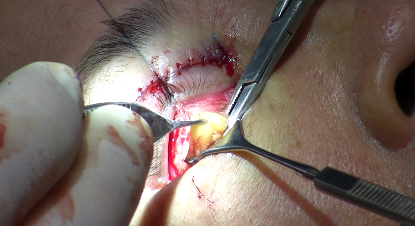
Repositioning works by releasing the herniated fat from its compartment, mobilising it downward over the inferior orbital rim, and anchoring it into the tear trough depression below. The fat that was causing the bag becomes the correction for the hollow that sits beneath it. The result, when well executed, is a smooth transition from lower lid to cheek - no bulge, no crater.
Pure fat removal is not obsolete. Patients with genuinely excessive fat volume and good skin quality may do well with careful targeted excision. The operative word is careful - under-excision leaves residual bags, while over-excision produces the hollowing that defined the older results. Many surgeons today take a hybrid approach: reposition what can be repositioned into the tear trough, remove only what remains genuinely excessive after that redistribution.
How Surgeons Assess Lower Lid Safety Before Operating
A thorough lower lid consultation spends considerable time on risk assessment before any technique discussion. Several specific evaluations determine not only whether surgery is appropriate but what additional steps are needed to perform it safely.
The Snap Test
The surgeon pulls the lower lid gently away from the eye and releases it, instructing the patient not to blink. In a lid with healthy muscle tone, it returns to position promptly - typically within about two seconds. In a lid with reduced tone, it returns slowly or requires a blink to reseat itself against the globe. Slow return - called poor snap - signals laxity that makes the lid more vulnerable to downward drift after surgery and is a meaningful predictor of complication risk.
Negative Vector Anatomy
Viewed in profile, the relationship between the cheekbone and the front surface of the eyeball matters. When the globe protrudes forward past the cheekbone - a configuration called negative vector - the lower lid has less bony support beneath it. This is an independent risk factor for lid retraction after surgery, identifiable on pre-operative photographs and lateral profile views. Patients with negative vector anatomy are not necessarily excluded from surgery, but their surgeon should account for it explicitly in the approach.
Scleral Show
The white of the eye - the sclera - should not be visible below the iris in normal forward gaze. If it is visible before surgery, that alone signals reduced lower lid tone and a higher baseline risk for the procedure. It can also appear as a new post-operative finding, indicating that the lid has drifted or been pulled downward - which is why a surgeon documents its presence or absence before operating.
When Laxity Is Found
If the snap test, vector assessment, or scleral show suggests laxity, the surgeon faces a decision about how to compensate. The options are not simply "operate or don't."
- Canthopexy tightens the lateral canthal tendon without detaching it - a less invasive addition suited to mild laxity, often added as a precaution even when laxity is borderline.
- Canthoplasty fully detaches and re-anchors the canthal tendon - a more robust correction for moderate to significant laxity, more involved to perform but more durable in its support.
- Both procedures are frequently added prophylactically at the time of blepharoplasty rather than waiting to see whether a problem develops afterward.
The Risk of Ectropion and Lid Retraction: What Patients Actually Need to Know
Ectropion is the outward rolling of the lower lid margin away from the eyeball. The lid pulls downward and outward, exposing the inner conjunctival surface, causing the eye to water, redden, and feel persistently irritated. In pronounced cases it disrupts vision. It is the complication most surgeons are most cautious about - and most forthright surgeons will discuss it in detail during consultation.
The mechanism is scar contracture. Healing tissue beneath the lid skin shortens over weeks and tugs the lid margin away from the globe. The subciliary approach carries a higher risk of this outcome because the incision sits precisely in the layer that contracts. The transconjunctival approach, which leaves external tissue undisturbed, carries a lower but not zero risk - particularly in patients whose baseline anatomy already puts them at higher starting risk.
The Treatment Ladder When Ectropion Occurs
Mild to moderate cases do not go straight to reoperation. The standard management moves through conservative measures first, because many early cases of post-operative lid retraction improve with time and deliberate intervention.
- Massage along the lower lid - started with surgeon approval in the early weeks after surgery - applies gentle upward pressure to counteract early scar tightening before it becomes fixed.
- Taping the lower lid upward at night maintains position and applies counter-traction while the scar matures.
- Lubricating eye drops manage the dry, exposed surface during this period and protect the cornea.
- If conservative measures have not resolved the problem after several months - published literature commonly cites around three to six months as the typical window - surgical revision is considered, typically involving scar release and, if needed, a spacer graft (such as cartilage or other tissue) to provide lasting lid support.
Rushing to revise an ectropion creates additional scar tissue in an area that has already been disrupted. The waiting period is not inaction - it is protocol.
Recovery Week by Week: What the First Eight Weeks Look Like
Lower lid recovery is substantially slower and less linear than upper lid recovery. Patients who have had upper blepharoplasty and expect a similar course are consistently surprised by how much longer the lower lid takes to settle.
The First Two Weeks
Swelling peaks in the first few days and is typically worst on the second or third morning. Bruising spreads downward across the cheek and may extend further than expected. Chemosis - swelling of the conjunctiva itself, making the white of the eye look gelatinous or puffy - is common and alarms patients who are not warned about it. It is almost always temporary. Cold compresses, sleeping with the head elevated, and avoiding anything that raises blood pressure - including strenuous exercise and alcohol - all help manage early swelling.
Weeks Three to Eight
Swelling improves week by week but does not resolve in a straight line. Many patients notice that one side clears faster than the other. Surgeon-supervised massage can often begin in the first two to three weeks after surgery, though the exact timing depends on individual healing and surgeon protocol - always follow your own surgeon's instructions. It helps prevent scar contraction and encourages residual fluid to disperse. The lower lid's slower recovery reflects its anatomy directly - less structural tissue means less support for fluid drainage, and lymphatic clearance in this region is genuinely less efficient than above. Significant swelling can persist for two to three months in some cases, noticeably longer than after upper blepharoplasty.
The Result at Six Months and Beyond
What patients see at six weeks is not the final result. Residual firmness, subtle asymmetries, and minor skin texture irregularities continue to evolve. Most surgeons expect significant improvement to be clearly visible by around three to six months. Scar maturation and the last traces of residual swelling can take longer - up to a year in some cases - which is worth keeping in mind before drawing conclusions from an early follow-up visit.
When Lower Blepharoplasty Is Not the Answer
Surgery is not the right intervention for every complaint about the under-eye area. Several presentations are better served by a different treatment, or will not be improved by standard lower blepharoplasty at all.
- Pure tear trough hollowing without fat herniation - hyaluronic acid filler placed carefully by an experienced injector is the appropriate treatment. Operating on an area that needs volume added, not fat managed, creates risk without addressing the anatomy.
- Festoons - chronic folds of skin and soft tissue that drape across the cheekbone below the lower lid. Festoons are structurally distinct from orbital fat herniation and are not corrected by standard lower blepharoplasty. Patients who have festoons frequently discover this only after surgery: the bags are resolved but the cheek folds remain, because the two problems require different interventions entirely. Surgeons who do not distinguish them at consultation leave patients unprepared for an incomplete result.
- Skin-only laxity without a significant fat component - fine lines, crepey texture, and loose surface skin are not caused by fat herniation. Fat-directed surgery does not address them. Laser resurfacing or a targeted skin-pinch procedure is a more appropriate approach for this presentation.
- Patients with significant lid laxity and no plan to address it - operating on an unsupported lower lid without adding canthal stabilisation is a preventable setup for complications. If the snap test shows significant laxity and the surgeon does not intend to address it, that is a specific question worth raising before signing a consent form.
- Existing filler in the tear trough area - patients who have previously had filler placed may find the fat herniation is worsening the overall appearance despite the filler. The filler is adding volume to an area already under pressure from herniated fat. This history needs to be disclosed at consultation and may affect timing or technique.
Questions to Bring to Your Consultation
A thorough surgeon addresses most of this unprompted. These questions surface the specifics and give you a basis for comparing how different surgeons approach the same anatomy.
- Which incision approach do you plan to use for my anatomy, and what is the reasoning?
- What did the snap test show, and do I need a canthopexy or canthoplasty?
- Are you planning fat repositioning, fat removal, or both - and why for my specific case?
- Do I have any festoons, and will this surgery address them?
- What is your personal rate of post-operative lid retraction or ectropion, and how have you managed those cases when they occurred?
- I have had filler in this area previously - does that affect your planning or the timing of surgery?
A surgeon who gives specific, considered answers to these questions - not reassurances, but actual answers grounded in your anatomy - is one who understands the complexity of what lower blepharoplasty requires. Vague confidence is not the same thing.
Frequently Asked Questions
Can I have lower blepharoplasty if I have already had filler in my tear trough?
Yes, but the history matters. Prior filler needs to be disclosed, and depending on how recently it was placed and how much was used, your surgeon may want to allow it to dissolve or may recommend hyaluronidase to clear it before proceeding. Filler on top of herniated fat complicates both the visual assessment of the problem and the surgical approach.
Is lower blepharoplasty always done under general anaesthesia?
Not always. The procedure can be performed under local anaesthesia with intravenous sedation as well as under general anaesthesia, and both are routinely used as day-case procedures. The choice depends on the complexity of the planned surgery, whether it is being combined with other procedures, and the patient's and surgeon's preference. Your anaesthetic options should be discussed explicitly at your pre-operative assessment.
How do I know if my problem is bags, a tear trough hollow, or both?
A rounded, protuberant bulge below the eye that you can see from across the room in normal lighting is characteristic of fat herniation. A shadowed groove running from the inner corner of the eye along the cheek - often more visible in certain lighting - points more to a hollow. Many patients have both: the fat bag at the lid and a hollow beneath it where volume has been lost. A surgeon will assess the two separately, and the distinction determines whether you need surgery, filler, or a combination.
What does ectropion actually feel like, and how would I know if it was developing?
Early lid retraction feels like persistent eye watering, a foreign body sensation, or dryness that does not respond normally to eye drops. Visually, you might notice the lower lid margin sitting lower than before surgery, or a small strip of white sclera becoming visible below the iris when looking straight ahead. Any of these signs in the weeks after lower blepharoplasty should prompt a call to your surgeon - early intervention with massage and taping is far more effective when started promptly.
Will lower blepharoplasty also help with loose, crepey skin under my eyes?
Not directly, if the approach used is transconjunctival - that route does not address skin at all. If skin laxity is part of your concern, it needs to be part of the plan: either a subciliary approach with skin excision, a separate skin-only pinch excision, or laser resurfacing to the surface. Clarify this at consultation, because patients sometimes assume the surgery will address more than the planned technique actually covers.
Why does swelling last so much longer after lower lid surgery than upper?
The lower lid sits in a region with less structural tissue to support it and slower lymphatic drainage compared to the upper. Gravity also works against it - fluid that accumulates in the lower lid has nowhere easy to drain. The result is that swelling that clears in a week or two after upper blepharoplasty can persist for two to three months at the lower lid, even in uncomplicated cases. This is a normal feature of lower lid anatomy, not a sign that something has gone wrong.
General information only, not medical advice. Individual anatomy and healing vary. See the disclaimer.