The yellow patches that appear near the inner corner of your eye are easy to dismiss. They are painless, flat, and slow-growing - easy to file away as cosmetic noise. But xanthelasma deserves more serious attention than its appearance suggests. It can signal abnormalities in cholesterol metabolism that a standard blood test will miss, and it has been independently linked to cardiovascular risk in people who have no idea anything is wrong. This guide covers what the deposits are, how to grade their severity, how each removal option performs over time, and what you can do after treatment to reduce the chance they come back.
What Xanthelasma Looks Like and Where It Appears
Xanthelasma palpebrarum presents as soft, flat plaques with a pale yellow or creamy-yellow color. They sit flush against the skin rather than protruding from it - not a bump you can feel under your fingertip, more of a subtle change in color and texture. The surface is smooth and waxy. The skin over the deposit moves freely, and the lesions are painless and non-itchy.
The most common starting point is the medial canthus - the inner corner - of the upper eyelid. As deposits develop over months to years, they can spread to the inner portion of the lower lid. In more severe cases, they extend across all four lids. Individual plaques range from just a few millimeters to well over a centimeter wide. They do not resolve on their own. Without treatment, xanthelasma persists and gradually enlarges.
Why Xanthelasma Forms
The deposits are collections of lipid-laden macrophages - immune cells that have taken up so much cholesterol and fat that they become what pathologists call foam cells. These accumulate in the dermis of the eyelid, producing the yellow color visible through the skin. The eyelid skin is the thinnest skin on the body, which is part of why lipid particles deposit here more readily than at other sites. Xanthelasma is most common in middle-aged adults and affects women more often than men.
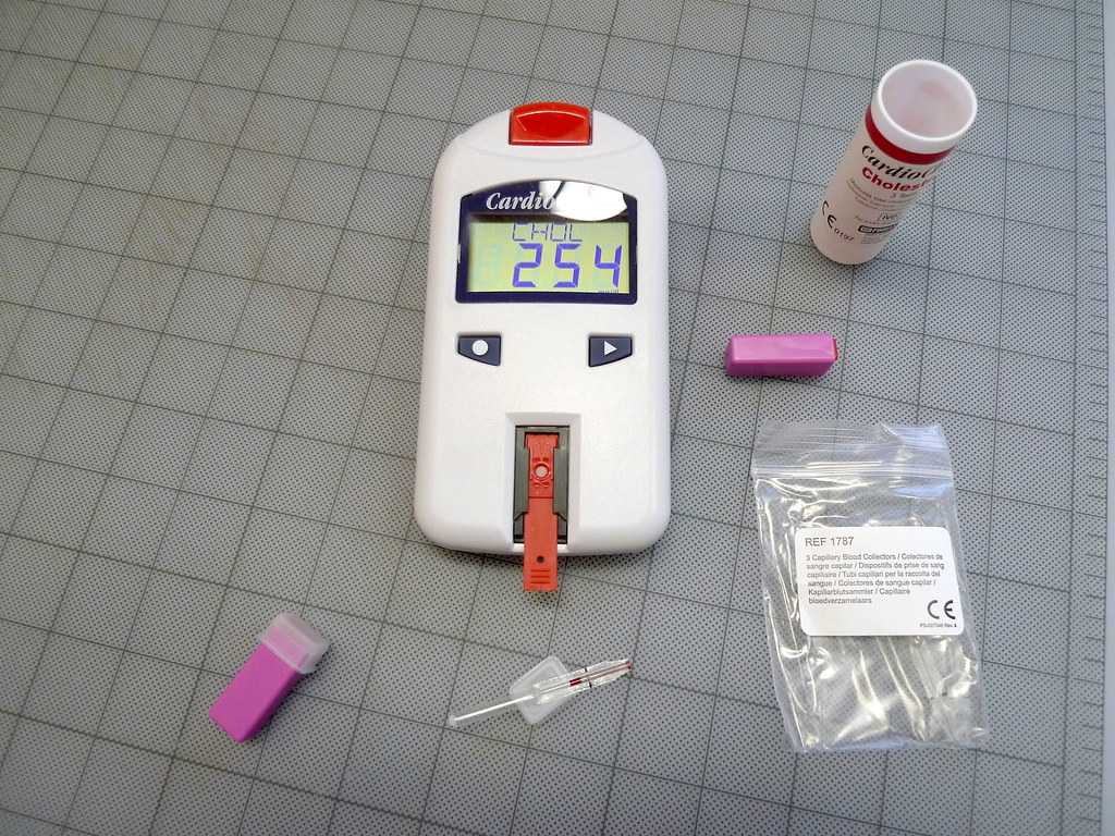
Here is what surprises most patients: approximately half of people diagnosed with xanthelasma have elevated cholesterol or triglycerides on a standard lipid panel. The other half have entirely normal numbers. Normal results do not rule out xanthelasma, and they do not rule out the cardiovascular implications that come with it.
The Heart-Disease Connection
Xanthelasma is an independent predictor of myocardial infarction, ischemic heart disease, and atherosclerosis. Independent means that the association holds even after accounting for conventional risk factors - age, blood pressure, smoking, and standard cholesterol levels. The American Academy of Ophthalmology has stated publicly that these apparently harmless-looking deposits near the eye may be a sign of heart disease, based in part on large prospective research linking xanthelasma to elevated cardiovascular event rates even in people with normal standard cholesterol.
That harmless bump near your eye may be a sign of heart disease.American Academy of Ophthalmology
The explanation for why this is true even in patients with normal cholesterol lies in what a standard lipid panel does not measure. People with xanthelasma tend to have raised apolipoprotein B, raised LDL particle number, and reduced levels of HDL subfraction 2 - the most cardioprotective component of HDL. A basic lipid panel measures total LDL cholesterol concentration and total HDL, but it does not distinguish between a person carrying a normal number of large, buoyant LDL particles and a person carrying an abnormally high number of small, dense, atherogenic ones.
Apolipoprotein B is worth knowing about because it directly counts the number of atherogenic particles in the blood rather than measuring their cholesterol content. Someone can have an acceptable LDL cholesterol number while their apolipoprotein B reveals a particle burden that is driving plaque formation. Xanthelasma appears to be a visible skin marker of that pattern.
A reasonable step after a xanthelasma diagnosis is a conversation with your GP about going beyond the standard panel - specifically asking about apolipoprotein B testing and a frank discussion of your full cardiovascular risk profile. Whether a cardiologist needs to be directly involved depends on age, family history, blood pressure, and other factors. That conversation belongs alongside whatever cosmetic treatment you pursue, not instead of it.
How to Tell It from Other Eyelid Bumps
Several common eyelid lesions get confused with xanthelasma. Getting the diagnosis right before treatment matters because each condition calls for a different approach.
- Milia - Tiny, hard, white cysts, typically 1 to 2 mm across, containing keratin rather than cholesterol. They feel firm under the finger rather than soft and waxy. Milia tend to appear in clusters across the cheeks and under the eyes and carry no link to lipid disorders or cardiovascular risk.
- Syringoma - Flesh-colored to slightly yellowish papules arising from sweat ducts. They appear in clusters just below the lower lash line, are more rounded and raised than the flat plaques of xanthelasma, and are not associated with cholesterol abnormalities.
- Sebaceous cysts - Larger, rounder, and often distinguished by a visible central pore at the surface. They fill with waxy sebum, feel firmer and more defined than xanthelasma, and are not linked to lipid metabolism.
In practice, xanthelasma is recognizable by three features together: the yellow-to-cream color, the flat plaque shape, and the medial upper-eyelid location. A physician can usually confirm the diagnosis on clinical inspection alone; biopsy is rarely needed. Knowing which type of bump you have before your consultation lets you describe it accurately and reach a treatment conversation faster.
The Four Grades and Why They Matter
Surgeons and dermatologists use a four-grade classification to describe how extensive the involvement is. Grade directly determines which treatment options are on the table and how complex the procedure needs to be.
- Grade I - Upper eyelids only. The most common presentation at first diagnosis, and the most straightforward to treat.
- Grade II - Upper lids plus the medial portion of the lower lids. Still localized to the inner canthal area, but requiring attention to both lids.
- Grade III - Medial upper and lower lids on both sides. The deposits are larger and bilateral, making surgical closure more demanding.
- Grade IV - Diffuse involvement extending laterally across all four lids. This presentation requires the most careful surgical planning and carries the highest recurrence risk regardless of method.
For Grade I and early Grade II lesions, a surgeon has genuine choices among excision, laser, and chemical peel. As grade increases, excision tends to become the preferred approach because non-surgical treatments cannot reliably reach the full depth of larger or thicker deposits.

Surgical Excision: The Gold-Standard Option
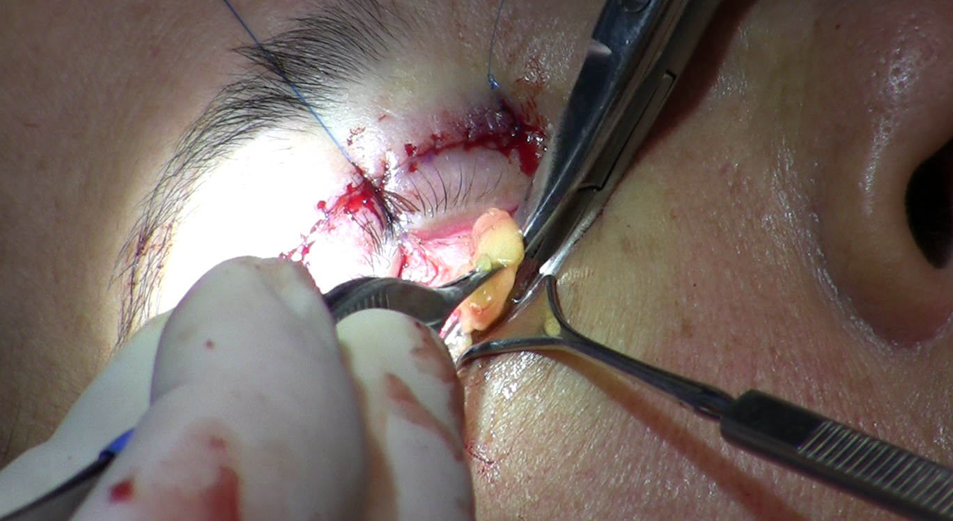
Surgery is the definitive first-line treatment for xanthelasma. The procedure is performed under local anesthesia as an outpatient visit and typically takes under 30 minutes. The surgeon maps the margins of the deposit, infiltrates local anesthetic, and removes the lesion with a scalpel or fine scissors, working to address the full depth of the accumulation.
Closing the wound in large lesions
For smaller deposits, the wound can usually be closed in a straight-line fashion without distorting the lid. The challenge comes with larger lesions. The eyelid has very limited redundant skin - there is not much to spare - so removing a wide area leaves a defect that cannot always be closed by simply pulling the wound edges together.
Surgeons handle this in two main ways. One is to plan the excision as an ellipse of skin using blepharoplasty technique - removing a matching arc of lid skin so the closure follows the same mechanics that prevent lid distortion in cosmetic eyelid procedures. The other, for the largest defects, is a skin graft, typically taken from the upper eyelid of the same patient, to fill the area without pulling the lid margin out of position. Asking which approach your surgeon plans for your specific lesion is a sensible question at any consultation.
Recovery and recurrence
Swelling and bruising around the eye are expected for one to two weeks. Most patients return to normal activity within that window. Recurrence following surgical excision runs at roughly 10 to 40 percent over a three-to-five-year period - a wide range, but also the lowest floor of any treatment method. Patients with Grade I or II disease who bring their lipid levels under control after surgery tend to land toward the lower end.
Laser Treatment: CO2 and Er:YAG
Ablative lasers vaporize tissue layer by layer. The surgeon passes the beam across the deposit repeatedly, removing thin laminae until the full depth has been addressed. This avoids a sutured incision and works well for deposits that are not too thick or wide.
CO2 laser
The carbon dioxide laser is the more powerful option. Published studies report high clearance rates in the majority of treated lesions, though outcomes vary by lesion grade, skin type, and length of follow-up; results in clinical series range from very high clearance in lower-grade lesions to more variable outcomes in higher-grade or multiply recurrent cases. The trade-off is a longer recovery: treated skin remains red and tender for several weeks as it re-epithelializes, and there is a meaningful risk of post-inflammatory hyperpigmentation, particularly in darker skin tones.
Er:YAG laser
The erbium laser achieves high clearance rates in most patients, with published studies reporting good to excellent outcomes in the majority of treated lesions, though results vary across series and depend on lesion characteristics and skin type. For patients who need to return to professional or social settings within a week or two, the erbium option is worth a direct conversation. The reduced thermal effect means less collateral heat spread to surrounding tissue, which is why recovery is faster.
For both laser options, published recurrence rates vary substantially across studies and patient populations, ranging from under 15 percent in some recent series to over 50 percent in others, depending on lesion grade, skin type, and whether underlying lipid levels are addressed after treatment. This range is generally higher than the surgical excision floor, reflecting the fact that laser treatment can miss lipid at the deepest margin of a thick deposit. Ask your provider about realistic recurrence expectations for your specific lesion grade and skin type.
TCA Chemical Peel
Trichloroacetic acid at high concentration - typically in the 70 to 80 percent range - is a well-established in-office treatment that works by chemically coagulating the lipid-laden cells within the deposit. A physician applies the acid to the lesion with a fine applicator, producing a white frost on the skin surface that shows where the chemical has penetrated. The treated tissue then sloughs off over approximately a week, and new skin forms underneath.
Clinical studies have shown TCA to produce meaningful patient satisfaction and an acceptable cosmetic result in most patients. The practical difference from laser is that multiple sessions are typically required - the deposit often improves incrementally across treatments rather than clearing in one pass. For patients who prefer a gradual approach or for whom laser equipment is not accessible or cost-prohibitive, TCA is a legitimate option.
The recurrence picture with TCA is generally less favorable than surgical excision, with published studies reporting recurrence in a substantial proportion of patients within the first one to two years; exact rates vary by study population, lesion characteristics, and whether lipid levels are managed after treatment. Pigment change remains the most common side effect. The risk is manageable with good technique and appropriate patient selection, but it warrants a clear discussion before committing to the approach.
| Method | Best suited for | Recurrence risk | Recovery |
|---|---|---|---|
| Surgical excision | Larger, deeper, or Grade III-IV lesions | Lowest of the three | Bruising 1-2 weeks |
| CO2 laser | Small to medium deposits, thinner skin | Moderate; varies by grade and skin type | Redness several weeks |
| Er:YAG laser | Small to medium; faster recovery priority | Moderate; published rates vary - discuss with provider | Shorter than CO2 |
| TCA peel | Small, superficial deposits | Higher than excision; multiple sessions often needed | Sloughing over roughly a week per session |
Recurrence: Honest Expectations and What Patients Can Do
Xanthelasma comes back for a meaningful number of patients regardless of method. The rate varies - lowest for surgery, highest for chemical peel - but no treatment guarantees permanent clearance. Two factors drive recurrence more than any other: whether the underlying lipid abnormality is being treated, and how extensive the disease was at the time of treatment. Grade IV involvement across all four lids carries higher recurrence risk across every modality.
What you can actually do
Managing cholesterol through diet, exercise, and medication after treatment demonstrably reduces recurrence risk. This is not vague advice - the metabolic environment that produced the original deposits can produce new ones if it remains unchanged. Steps worth discussing with your physician include:
- Following up on any lipid abnormalities found in your workup, including apolipoprotein B if elevated
- Reducing dietary saturated fat and refined carbohydrates, which lowers both LDL particle number and triglycerides
- Regular aerobic exercise, which raises HDL cholesterol and improves HDL function
- Statin therapy or other lipid-lowering medication if your physician determines it is warranted based on your full cardiovascular risk profile
- Returning for a dermatologic or oculoplastic follow-up annually for at least three years after treatment - catching a recurrence early, when deposits are small, keeps the next treatment simpler
Choosing a Provider
Xanthelasma treatment is not a routine skin procedure. The lid margin must open and close precisely, the skin cannot be left too tight or too loose, and the lacrimal drainage system near the inner corner must be protected throughout. Errors in this area affect lid function and potentially vision, not just appearance.
Oculoplastic surgeons - formally called ophthalmic plastic and reconstructive surgeons - train specifically in eyelid and orbital surgery and are the most consistently qualified specialists for this work. Experienced dermatologic surgeons with a documented focus on periorbital procedures are also well-positioned. Some plastic surgeons with extensive facial and eyelid experience are appropriate as well, though the inner canthus and lid margin are not routine territory for general plastic surgery practice.
When you meet with a provider, ask these questions in order:
- How many xanthelasma procedures have you performed, and can you show before-and-after photographs specific to cases similar to mine?
- What grade do you assess my lesions to be, and how does that drive your recommendation?
- If surgical excision is the plan, how will you close the defect - primary closure, blepharoplasty technique, or a graft?
- What lipid workup do you recommend I complete before or alongside treatment, and should I bring my GP or a cardiologist into this conversation?
A provider who answers those questions specifically - with reference to your anatomy, your grade, and your medical picture - is demonstrating the kind of familiarity that periorbital work requires.
Frequently Asked Questions
Can xanthelasma resolve on its own without treatment?
No. Xanthelasma does not resolve spontaneously. The deposits are stable collections of lipid-laden cells that do not reabsorb once established. Even bringing cholesterol levels under control will not dissolve existing lesions - though it does reduce the chance of new ones forming after treatment.
My cholesterol test came back normal. Do I still need further cardiac evaluation?
Yes, a conversation with your doctor is still appropriate. Xanthelasma has been linked to cardiovascular events independently of standard cholesterol numbers, and is associated with qualitative lipid abnormalities - specifically raised apolipoprotein B and reduced HDL subfraction 2 - that a basic lipid panel does not capture. A more detailed workup gives a far more accurate picture of your actual risk.
Will statins or cholesterol medication shrink existing deposits?
Lipid-lowering medications do not reliably dissolve established xanthelasma. Their value is in changing the metabolic environment to reduce the likelihood of new deposits forming - making them a useful adjunct to treatment, not a standalone fix. Patients who control their lipid levels after removal do tend to have lower recurrence rates, which is a strong reason to pursue that discussion with your physician.
Is the Er:YAG laser better than CO2 for someone who cannot take extended time off?
For patients who need a shorter visible recovery window, Er:YAG has a practical advantage: its reduced thermal effect means less prolonged redness after treatment. Published clearance and recurrence data vary across studies, so the right choice depends on your specific lesion grade, skin tone, and provider expertise. If your lesions are small and superficial, the trade-off may be acceptable - worth discussing directly with your provider.
Is xanthelasma removal covered by health insurance?
In most cases, removal is classified as cosmetic and is not covered. An exception can sometimes be made when deposits are large enough to obstruct the visual field - a situation that can arise in Grade IV involvement - in which case a functional argument for coverage may apply. Check with your insurer and ask your surgeon whether formal documentation of visual field compromise is relevant to your case.
How soon can I expect to see recurrence after treatment, and what does it look like?
Early recurrence typically looks like a faint yellow tinge returning to the treated site, usually starting as a small area near the medial canthus. Published timelines and rates vary by method and individual patient factors; chemical peels tend to have the shortest recurrence-free intervals and laser and surgery progressively longer ones. Annual follow-up appointments make it possible to catch re-growth while it is still small and manageable.