Most procedure guides tell you that upper blepharoplasty removes excess skin and restores a more open eye. What they skip - the upright marking session, the two fat pads with different colours and different fates, the moment a surgeon finds a hidden lid droop inside yours - is exactly what patients want to know before consenting. This guide follows the operation from the first pen mark through the final stitch.
Why Surgery Starts Before the Table
The first surgical decision happens while you are sitting in a chair, not lying on an operating table. The surgeon draws the incision lines before anaesthesia, before you recline, before the face is draped. The reason is gravity.
When you sit upright, the weight of the forehead and brow pulls overhanging skin downward. Lying flat shifts that tissue back toward the scalp. A marking made supine records a lid position that simply does not exist when you walk out of the clinic - the incision would land in the wrong place, and the result would reflect that.
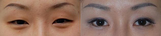
The crease is marked at a specific height above the lash margin. In Caucasian anatomy the target sits slightly higher in women than in men. In East Asian anatomy it is placed deliberately lower to respect the patient's natural crease rather than imposing a different one. These are structural decisions that determine whether the result looks appropriate to the person's face or conspicuously altered.
Deciding How Much Skin to Remove
Knowing where to cut is step one. Knowing the safe upper limit is what protects the patient from the operation's most serious risk: over-resection.
Surgeons use one of two methods - or both - to define that limit before the incision is made.
| Method | How it works | What it measures | Limitation |
|---|---|---|---|
| Caliper rule | Calipers confirm that a minimum margin of lid skin remains after any planned excision - ask your surgeon which threshold they apply | Absolute floor for full lid closure | Does not account for individual skin laxity or elasticity |
| Pinch test | Non-toothed forceps pinch at nasal, central, and lateral points until the lashes just begin to evert | Safe removal limit at three horizontal positions across the lid | Technique-dependent; requires consistent execution to standardise |
| Combined use | Both checks performed; the more conservative result governs what is marked | Redundant cross-check across different methods | Only meaningful when both steps are executed carefully |
When too much is removed, the eye cannot close completely - a condition called lagophthalmos. Mild cases resolve as post-operative swelling settles. Significant cases leave the cornea exposed during sleep, and that carries real risk to vision.
The Incision: Where It Sits and Why It Disappears
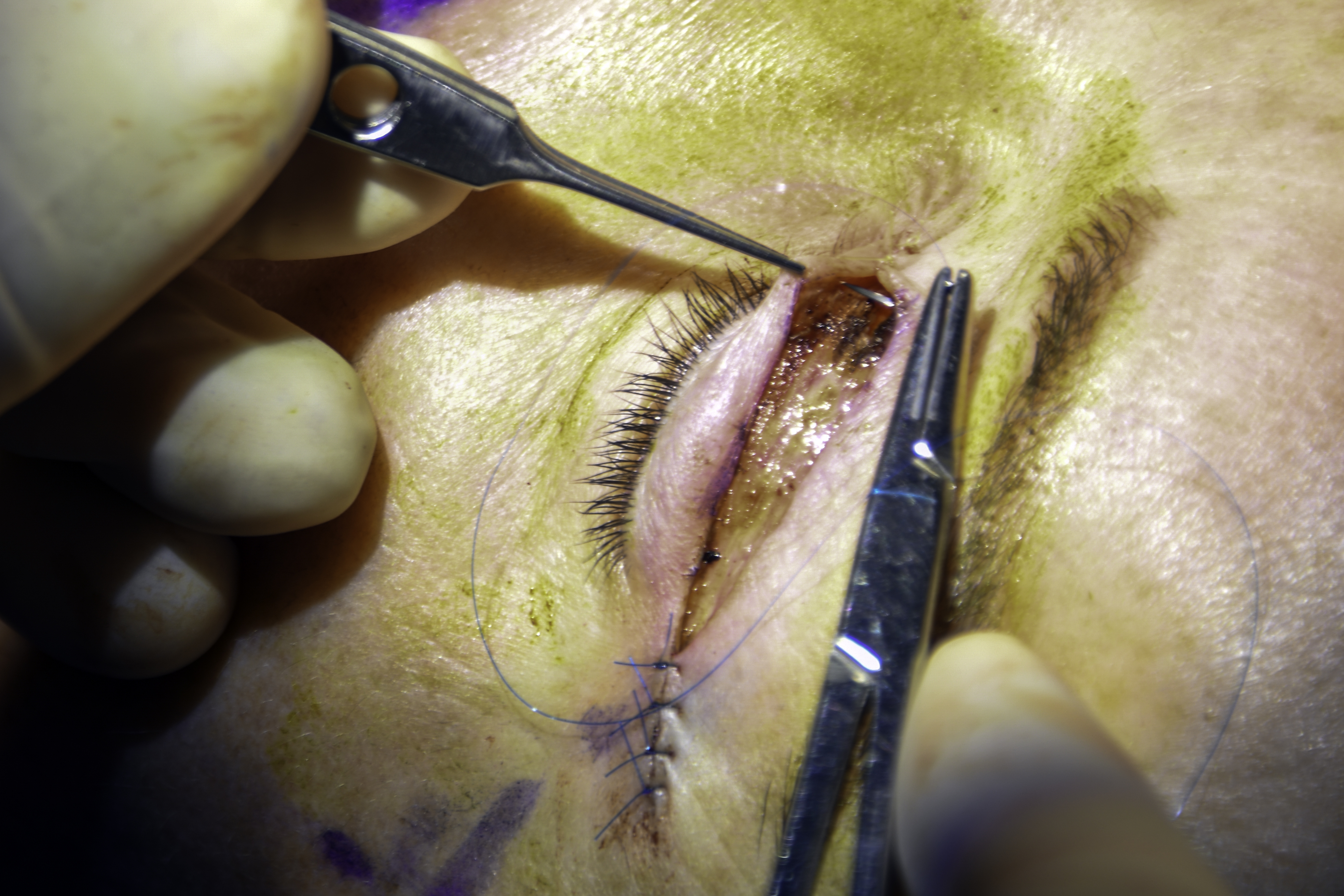
The incision is placed inside the pre-existing eyelid crease. This is not just for concealment - it is the anatomically correct location. The levator palpebrae superioris, the muscle that lifts the upper lid, sends fibres that insert into the skin at precisely this point. Every time the eye opens, the skin folds inward here. The healed scar sits within that fold and is not visible when the eye is open.
Upper eyelid skin is among the thinnest on the body, thickening progressively toward the brow. Thin skin closes with less tension, produces finer scar tissue, and remodels more completely than skin elsewhere on the face. This is why well-placed blepharoplasty incisions routinely become nearly undetectable within a few months.
Inside the Lid: Two Fat Pads, Two Different Fates
Once the skin is divided and the orbital septum opened, the surgeon finds two distinct fat pads. They look different from each other, and that difference shapes how each one is managed.
Identifying the pads
- Central fat pad (preaponeurotic): yellow, soft, and located directly behind the central septum in the mid-orbit
- Nasal fat pad (medial fat): white and more fibrous in texture, sitting in the inner corner of the orbit, separated from the central pad by the trochlea of the superior oblique muscle
A gentle tug on one pad will not move the other. That physical independence confirms which compartment is open and prevents the surgeon from inadvertently working in the wrong space.
Why the central fat is usually left alone
Older technique routinely removed both pads. The results, decades later, show what went wrong. Removing the central fat causes superior sulcus hollowing - a sunken, skeletonised appearance in the upper orbit that looks more aged than the fullness it was meant to correct. This is the hollow-eyed look associated with aggressive blepharoplasty of earlier eras. Modern technique treats the central fat as a volume asset and leaves it.
The nasal fat pad is the one most often responsible for visible upper lid fullness in the inner corner. When it needs addressing, the surgeon opens a small section of the medial orbital septum, expresses the pad gently, and sculpts it conservatively. The goal is reduction, not elimination.
The Orbicularis Muscle: Why It Stays
The orbicularis oculi is the ring of muscle just below the skin that controls the blink. In earlier blepharoplasty practice, a thin strip of it was routinely excised along with the skin to produce a sharper crease definition. Most surgeons have moved away from this.
The blink is not a passive movement. It depends on coordinated orbicularis contraction to spread the tear film and protect the corneal surface. Disrupting the muscle increases the risk of incomplete lid closure and corneal exposure, particularly during sleep. In patients with pre-existing dry eye disease, even partial orbicularis disruption can escalate a manageable condition into a symptomatic one requiring ongoing treatment.
Recent comparative studies have found no significant difference in cosmetic outcomes - scar quality, crease definition, or patient satisfaction - between cases where a muscle strip was excised and those where it was preserved. The evidence supports what many surgeons had already adopted in practice: leave the muscle and accept nothing in the cosmetic result.
The Hidden Ptosis Surprise
One of the more consequential moments in an upper blepharoplasty comes when the surgeon opens the lid and finds a levator aponeurosis that is stretched or partly detached from where it should be.
The levator aponeurosis is the broad, flat tendon that connects the levator muscle to the upper lid. With age it can loosen - a condition called aponeurotic ptosis. The lid droops not because of extra skin but because its internal support has weakened. Removing skin from a lid that is structurally drooping makes the ptosis more visible, not less. The eye looks sleepier after surgery than before.
Pre-operative examination catches obvious cases, but a stretched aponeurosis is sometimes only apparent once the lid is open on the table. When this happens, the blepharoplasty incision already in place provides direct access to the levator. The aponeurosis can be advanced and re-anchored through that single opening - no second incision, no second recovery period.
Signs worth raising at your pre-operative consultation:
- A high or poorly defined upper lid crease on one or both sides
- One lid that consistently sits lower than the other
- A lid that droops more visibly by the end of the day than in the morning
- Wider lid separation on downgaze than would normally be expected
Protecting the Levator: Where the Septum Is Cut
The orbital septum must be opened to reach the fat pads. The levator aponeurosis lies directly behind it. If the septum is incised at its lowest point, the blade works within millimetres of the levator. One careless pass causes post-operative ptosis - the lid drop that was not present before surgery.
To reduce that risk, surgeons incise the septum slightly above its lower border. This positions the entry point away from the levator attachment and creates a working margin before the tendon even comes into view. It is a small technical decision the patient never sees, and it directly explains the mechanism behind one of the procedure's most discussed complications. Post-operative ptosis following blepharoplasty is most often caused by accidental trauma to the aponeurosis at this specific step - not during the skin excision, not during fat sculpting, but here at the septum.
Closure and the First Week
Closing the incision does more than bring skin edges together. The sutures also re-establish the eyelid crease. A running or interrupted suture along the incision line incorporates small bites of levator fascia on each pass, anchoring the overlying skin to the same layer where the natural fold forms. This anchoring produces the defined crease that persists after healing.
The closing sequence, in order:
- Any fat sculpting and levator work is confirmed complete; bleeding is controlled thoroughly
- Skin edges are approximated and checked for symmetry before any sutures are placed
- The suture line is placed incorporating levator fascia at intervals to set the crease
- Closure tension is checked - the patient should be able to gently close the eye without strain
- Cold compresses are applied as the patient moves to recovery
Non-absorbable sutures come out at day 5-7 in the clinic. Comparative studies have found equivalent cosmetic outcomes between non-absorbable and absorbable alternatives; the choice reflects surgeon preference and patient tolerance. At the point of removal, the incision line looks red and linear, and the crease appears exaggerated by swelling.
What the first week typically brings:
- Bruising extending to the upper cheek, usually peaking around day three before gradually clearing
- Swelling that is worst in the morning and improves through the day as lymphatic drainage works
- Light sensitivity and mild blurring from lubricating eye drops used to protect the corneal surface
- A short clinic visit for suture removal at the end of the week, generally low in discomfort
Most swelling resolves within two to three weeks. Final scar softening and colour fading continue over three to six months.
How Long the Result Lasts - and What Eventually Undoes It
Upper blepharoplasty results typically persist for around a decade or more in many patients before visible changes return, though the range varies considerably depending on age at surgery, skin quality, and how quickly brow descent progresses. But the mechanism behind that return is not what most patients expect.
The skin removed during surgery does not grow back. What happens is that the forehead and brow continue to descend with age - driven by soft tissue volume loss and the sustained effect of gravity on facial structures that were already moving before surgery. When the brow drops, it shortens the distance between itself and the upper lid, and the overhanging skin reappears. It is brow descent pushing down from above, not new skin growing from below.
The longevity of a blepharoplasty result depends less on the operation itself than on how quickly the brow and forehead continue to descend after it.
This is why brow position is assessed before every upper blepharoplasty consultation. A patient whose brow has already descended significantly may find that addressing only the lid produces a shorter-lived result. It is also why lower blepharoplasty results tend to outlast upper ones: the fat removed from the lower orbit does not regenerate, and there is no equivalent structure above the lower lid to push tissue back into view over time.
General information only, not medical advice. Individual anatomy and healing vary. See the disclaimer.