Small white bumps appear along a blepharoplasty incision 4 to 6 weeks after surgery, often after patients have concluded the recovery is going well. These are milia - tiny keratin cysts, not pimples, not infections, not a sign of surgical error. They are the single most commonly reported minor complication of upper eyelid surgery. They are also highly treatable, and most resolve without any procedure at all. What patients need is an explanation of what exactly they are looking at, how to tell a milium from something more serious, and what the decision to treat actually involves.
What Milia Actually Are: Tiny Keratin Cysts, Not Pimples or Whiteheads
A milium is a sealed, self-contained keratin cyst sitting just beneath the skin surface. Its wall is lined by stratified squamous epithelium - the same layered cells that form normal outer skin. The interior holds tightly packed lamellar keratin, the fibrous protein found in skin, hair, and nails. There is no bacteria, no pus, and no infection inside the cyst. Nothing about it is related to acne or blocked pores in the conventional sense.
That sealed structure explains why milia resist squeezing. Without breaching the cyst roof, the keratin contents have no exit path. Each bump is firm and dome-shaped, typically 1 to 2 mm across, white or yellowish. It is non-tender and non-inflamed. Pressing it gently feels like a small bead under the skin. These physical characteristics are the first thing that distinguishes a milium from every other post-blepharoplasty bump that might appear along the same incision line.
Post-surgical milia are classified as secondary milia, which separates them from primary milia that arise spontaneously on the face without any triggering injury. Both types are histologically identical - lined by stratified squamous epithelium and filled with lamellar keratin. The difference is origin only, and that origin matters because it explains precisely why a blepharoplasty incision creates them.
Why Wound Healing Produces Them: The Secondary Milia Mechanism
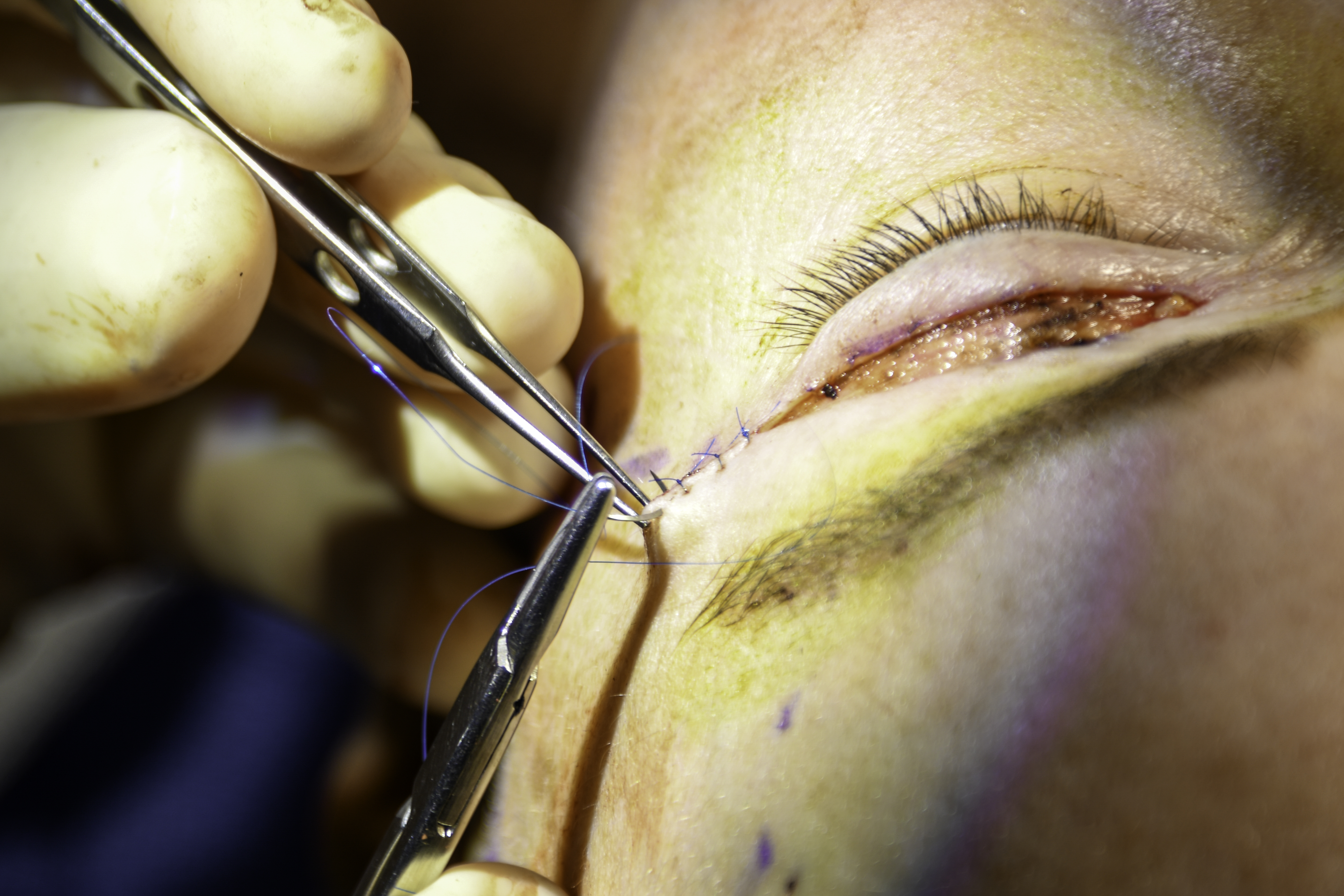
The upper eyelid crease contains a high density of adnexal structures: eccrine sweat glands with their surface ducts, hair follicles, and associated sebaceous glands. A blepharoplasty incision cuts through this tissue. During healing, regenerating epithelial cells resurface the wound. Some of those cells - along with remnants of eccrine duct walls and follicular fragments that were severed during surgery - become enclosed within the dermis rather than migrating to the skin surface.
Once sealed inside the dermis, those cells continue doing what epithelial cells do. They produce keratin. The keratin accumulates because there is no exit route. The result is a small epithelial-lined pocket that fills progressively until it is visible from the surface. That is the secondary milia mechanism in full: disrupted adnexal structures, enclosed during wound repair, build a keratin sac.
This is not a foreign body reaction and it is not an infection. It is a normal cellular process occurring in the wrong anatomical location because incision and healing redirected the tissue architecture. Primary milia arise from the follicular infundibulum without surgery. Secondary milia arise because surgery interrupted the normal exit pathways for keratin-producing cells and enclosed them where they could not drain.
When They Appear, How Common They Are, and Why Suture Choice Changes the Odds
Milia do not appear in the first days after surgery, when bruising and suture tracks are expected. They emerge during the active remodeling phase of healing, typically 3 to 8 weeks post-operatively - often after suture removal and after patients have assumed the recovery is finished. That late timing is what makes them so alarming. New bumps at week 5 or 6, after everything seemed fine, feel like something going wrong. They are not.
Multiple published complication reviews identify milia as the most common minor complication of upper blepharoplasty. Incidence varies considerably, and one of the factors with a directly measurable effect is suture technique. A comparative study of suture types in upper blepharoplasty (PMC8874258) found milia rates of roughly 2% with fast-absorbing gut sutures versus approximately 17% with running cutaneous locked Prolene sutures. A 2024 systematic review comparing absorbable and non-absorbable sutures (PMC12505724) found no clinically significant difference in functional outcomes or scarring, but noted that certain non-absorbable suture styles carry a meaningfully higher milia burden - a finding that has increasingly shifted some surgeons toward fast-absorbing gut for upper lid crease closure.
Post-operative wound care may also play a role. Heavy occlusive ointments applied beyond the first week of healing can trap keratin debris near the surface and contribute to cyst formation. Transitioning to a lighter product after initial healing, on your surgeon's guidance, is a reasonable step.
What Milia Look Like and How to Tell Them Apart From Other Post-Blepharoplasty Bumps
Accurate identification matters because the treatment for milia, suture granulomas, and inclusion cysts is different in each case. Attempting needle extraction on a suture granuloma, for example, does nothing useful and delays the correct treatment.
| Lesion | Size | Appearance | Tender? | Treatment |
|---|---|---|---|---|
| Milium | 1-2 mm | White or yellowish dome, no surrounding redness | No | Observation, needle deroofing, or topical tretinoin |
| Suture granuloma | Variable | Red, raised, may show a thread end at the surface | Yes - often marked | Corticosteroid injection or suture fragment removal |
| Inclusion cyst (epidermoid) | 3 mm or larger | Skin-colored or white, prominent wall, slow growth over months | No unless secondarily inflamed | Elliptical excision to remove the entire cyst wall |
| Stitch-track reaction | Punctate, follows suture path | Small crusted dots or mild pitting along the incision line | Minimal | Usually resolves without treatment |
Signs you can assess yourself
- A milium is firm, round, and dome-shaped with no surrounding redness - pressing it gently feels like a tiny bead just under the skin surface
- A suture granuloma is inflamed and tender - the skin around it is pink or red, and the bump itself is sore to touch; a visible thread end at the surface confirms retained suture material
- An inclusion cyst is noticeably larger than a milium, grows slowly over weeks to months, and has a more prominent raised contour - needle deroofing will not adequately treat it because the cyst wall must be surgically excised to prevent recurrence
- A stitch-track reaction follows the exact path of the suture line in a dotted or pitted pattern and fades on its own as skin resurfaces
Any bump that is tender, rapidly growing, or surrounded by spreading redness and warmth warrants a call to your surgeon - those features are inconsistent with milia and need clinical assessment.
Upper vs. Lower Eyelid: Why the Crease Incision Gets More Milia
Milia after blepharoplasty occur predominantly along upper eyelid incisions. Lower blepharoplasty performed via the transconjunctival approach - where the incision sits on the inner surface of the eyelid and the outer skin is never cut - produces almost no milia, because the mechanism depends on disrupted skin-surface adnexal structures and those structures are never entered. Even the subciliary approach used for lower lid skin removal produces milia less often than upper crease incisions do.
Two anatomical factors explain the upper lid's higher rate. First, the skin of the upper eyelid crease is among the thinnest skin on the human body, which means adnexal structures are packed into a compact layer. Any incision through that layer disrupts more eccrine duct openings per millimeter than a comparable incision on thicker facial skin. Second, the upper lid crease has a higher density of eccrine sweat glands than the lower lid - which means more potential sources for cyst formation when ducts are severed and enclosed during healing. The incision is placed precisely where gland density is highest.
Watch and Wait: When Doing Nothing Is the Right Choice
Most milia resolve spontaneously. As the incision matures, the thin skin overlying each cyst gradually thins further and eventually ruptures naturally, releasing the keratin contents without any intervention. This typically takes 8 to 12 weeks from when the milia first appear. For many patients, the full timeline from surgery to resolution without treatment is 3 to 4 months.
Signs that observation is the right call
- Bumps are 1-2 mm, white or yellowish, and completely non-tender
- The count has not grown substantially over 2 to 3 weeks of monitoring
- There is no surrounding redness, warmth, or swelling
- The incision line itself is healing cleanly without signs of separation or infection
Surgeons generally recommend waiting at least 6 to 8 weeks after milia appear before considering any procedure, because a lesion already resolving on its own needs no intervention. Taking a weekly photo under consistent lighting is a practical way to track whether bumps are fading, stable, or multiplying - that information makes the watch-versus-treat conversation at a follow-up appointment straightforward.
In-Office Removal: Needle Deroofing, Electrodesiccation, and CO2 Laser
When milia persist beyond 12 weeks, continue to multiply, or cause enough cosmetic concern that the patient does not want to wait further, in-office removal is straightforward. The choice of method depends on how many milia are present and whether prior extraction attempts have already been made.
Needle deroofing: the standard approach
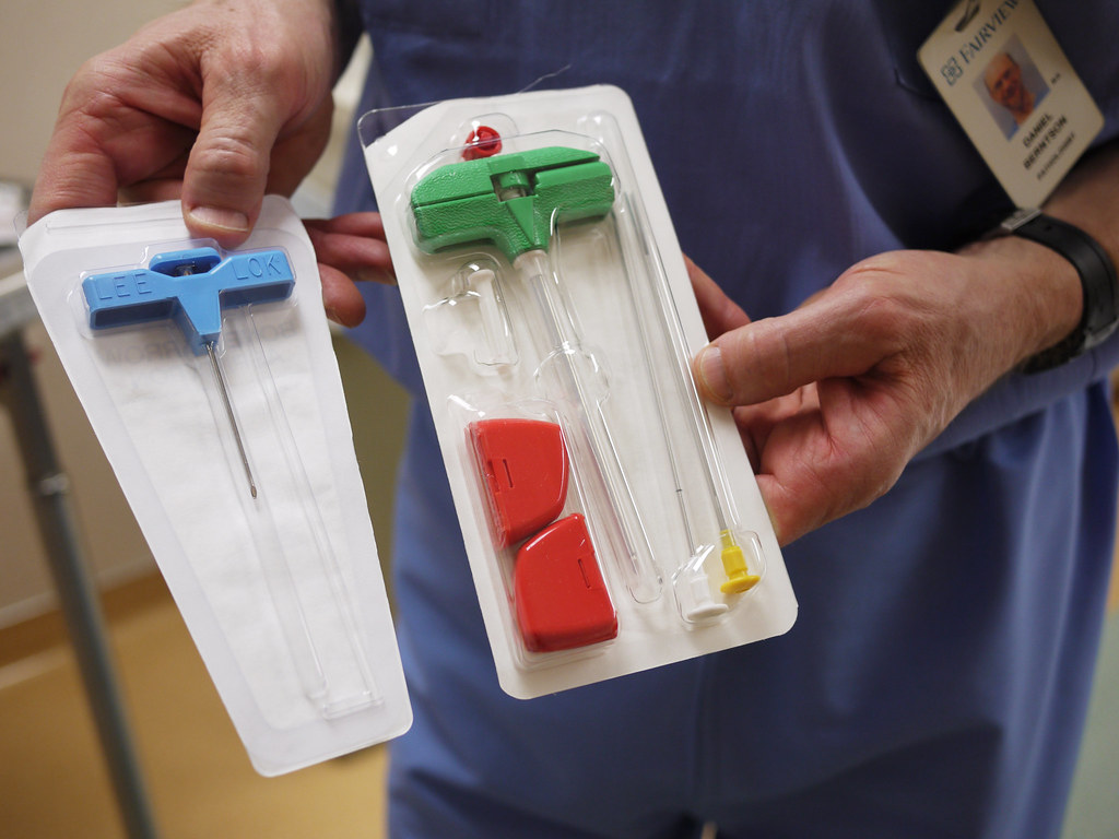
This is the first-line technique for isolated or small clusters of milia. The provider uses a sterile No. 11 blade tip or a fine-gauge hypodermic needle to make a shallow nick in the skin directly over the cyst dome. Opening the roof correctly is the step that matters - once it is breached at the right plane, the keratin core expresses easily with a comedone extractor or light finger pressure, coming out as a firm, pearlescent plug. The procedure takes seconds per milium. No sutures are needed, and downtime is minimal. The treated spot may appear slightly pink for a day or two.
The procedure in sequence:
- Cleanse the eyelid skin gently with a mild antiseptic
- Apply topical anesthetic cream if needed and allow adequate dwell time
- Use a sterile No. 11 blade or fine-gauge needle to nick the skin directly over the cyst roof with a single shallow pass
- Apply gentle pressure with a comedone extractor to express the keratin plug cleanly
- Cleanse the area and apply a small amount of antibiotic ointment
Electrodesiccation and CO2 laser
For clustered milia - several lesions grouped closely together - or for bumps that did not respond to needle deroofing, electrodesiccation uses a low-level electrical current to break down the cyst wall. It is faster than individual extraction when many lesions need treatment at one visit. CO2 laser ablation addresses the same scenario with a different energy source. Both produce a small treated zone that heals over 1 to 2 weeks with temporary local pinkness. On thin eyelid skin, the clinician adjusts settings carefully to avoid deeper thermal involvement.
Topical Tretinoin for Persistent Milia: How to Use It Near the Eye Without Damaging the Incision
Tretinoin accelerates epidermal turnover and thins the cornified layer, which can gradually thin the skin overlying a milium and allow the keratin to expel without any physical extraction. This makes it a reasonable option for patients with multiple persistent milia who prefer a topical approach, or for those whose surgeon wants to minimize in-office visits.
The periorbital skin is considerably more sensitive to retinoid irritation than the cheeks or forehead. Applied at standard facial concentrations to healing blepharoplasty scarring, tretinoin can cause significant redness, peeling, and discomfort. The approach requires restraint:
- Start at the lowest available strength, applied at night only
- Target only the milia themselves - not a broad coating across the eyelid skin
- Use the smallest amount that covers the area; thin eyelid skin absorbs topical products readily
- Expect 4 to 8 weeks for meaningful reduction - not days
- Stop and contact your surgeon if notable redness, peeling, or discomfort develops along the incision line
Tretinoin on a healing blepharoplasty incision requires your surgeon's explicit clearance - the same product used safely on intact cheek skin can disrupt wound remodeling when applied too early or too broadly on thin eyelid tissue.
Most surgeons will not recommend tretinoin near the incision until at least 6 to 8 weeks post-operatively and many prefer waiting until 12 weeks before introducing any active ingredient to that area. Do not begin on your own initiative based on general skincare guidance.
What Not to Do: Why Squeezing Without Opening the Roof Makes Things Worse
The instinct when seeing a white bump is to squeeze it. For a milium on a blepharoplasty incision, that instinct creates a worse problem than the original cyst.
A milium is sealed. Squeezing without first nicking the roof does not open the roof - it drives the keratin contents sideways and downward into the surrounding dermis. The dermis is not designed to contain loose keratin. The immune system treats displaced keratin as a foreign body and launches an inflammatory response. That response can produce a larger, more prominent bump than the original milium, trigger post-inflammatory hyperpigmentation on already-thin eyelid skin, or create the conditions for a true epidermoid inclusion cyst - which is larger, grows over months, and requires surgical excision with the entire cyst wall removed rather than a simple needle procedure.
Post-inflammatory hyperpigmentation along the eyelid incision is harder to address than the original milia. Thin eyelid skin does not respond as well to the bleaching agents and resurfacing lasers that work on thicker facial skin, and the discoloration can persist for months. The risk is not worth it when a brief in-office procedure or a waiting period accomplishes the same result cleanly.
Frequently Asked Questions
How do I know if the bumps on my eyelid incision are milia and not an infection?
Milia are firm, non-tender, and have no surrounding redness or warmth. An infection presents with redness spreading around the bump, warmth, tenderness on contact, and sometimes discharge. If a bump along your incision hurts when pressed, is surrounded by spreading redness, or you develop fever or increased swelling in the eyelid, contact your surgeon the same day rather than assuming it is a milium.
My surgeon used dissolvable sutures - can I still get milia?
Yes, though the risk is lower. Published data shows fast-absorbing gut sutures carry a substantially lower milia rate than certain non-absorbable running locked techniques, but no suture type eliminates the risk entirely. Milia form from disrupted adnexal structures during wound healing, which happens regardless of suture material - the suture choice influences the degree of tissue reaction, not the underlying mechanism.
How long should I wait before asking my surgeon to remove milia?
Most surgeons recommend waiting at least 8 to 12 weeks after milia appear, since many clear on their own within that window. If bumps are stable or increasing at 12 weeks, or if they are causing significant concern, that is a reasonable time to discuss needle deroofing. There is no medical urgency to treat sooner - early removal simply removes lesions that might have resolved without any procedure.
Will milia leave a permanent mark on my eyelid scar?
Milia that resolve spontaneously leave no visible mark - the keratin plug exits through naturally thinning skin without trauma to the surrounding tissue. Needle deroofing, performed correctly by an experienced clinician, is also unlikely to leave a scar. The scenario most likely to result in lasting discoloration is repeated home squeezing, which drives keratin into the dermis and triggers inflammation on skin that is already thin and sensitive.
Can I use an over-the-counter retinol product instead of prescription tretinoin?
Over-the-counter retinol converts to retinoic acid on the skin at a lower effective rate than prescription tretinoin, which makes it gentler - but it also means slower and less predictable results for milia. More importantly, the timing and application guidance is the same: do not apply any retinoid product near a blepharoplasty incision without your surgeon's clearance, regardless of whether it is prescription strength or over the counter. The thin, healing skin around the incision behaves differently than the intact facial skin these products are formulated for.
Is milia formation a sign that my surgeon made a technical error?
No. Milia are a product of normal wound healing in skin with a high density of adnexal structures - they occur even with meticulous technique. Suture choice influences incidence, and surgeons aware of that data increasingly favor lower-milia-rate techniques, but some rate of milia formation is an expected feature of upper eyelid surgery rather than a complication in the sense that scarring or wound dehiscence would be. Their appearance does not indicate that the procedure was performed incorrectly.
General information only, not medical advice. Individual anatomy and healing vary. See the disclaimer.