Blepharoplasty is one of the most commonly performed cosmetic procedures, and the overwhelming majority of patients recover without serious incident. But within the informed-consent process sits a complication that deserves a plain-language explanation rather than a single line of fine print: retrobulbar hemorrhage, or bleeding behind the eye. Its danger has almost nothing to do with the amount of blood involved and everything to do with anatomy. Understanding the mechanism - and the specific symptoms that signal it - is the difference between a close call and a permanent outcome.
What "Retrobulbar Hemorrhage" Actually Means
Retrobulbar means behind the globe - behind your eyeball. The eye sits inside a bony socket called the orbit, enclosed on four sides by flat bones of the skull. At the back, the optic nerve exits through a narrow channel. The only direction with meaningful give is forward, through the eyelids.
Blepharoplasty, whether on the upper or lower lids, requires the surgeon to work near the orbital fat: cushioning tissue that fills the space between the eyeball and the bony walls. Small blood vessels thread through this fat. If one bleeds after surgery, the blood has almost nowhere to go. A bruise on your cheek disperses outward through loose skin. Blood behind the eye is hemmed in by bone on every side. It can only push in one direction - forward, against the back of the globe.
The term hemorrhage implies a dramatic rupture. In practice, even a modest accumulation of blood in a rigid closed space can generate enough pressure to matter. Size of the bleed is secondary to the pressure it creates.
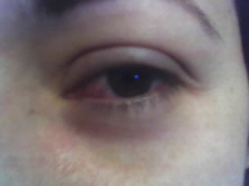
How Common Is Vision Loss After Blepharoplasty
The published numbers are specific and worth knowing directly, because "extremely rare" conveys nothing about actual risk.
Orbital hemorrhage after blepharoplasty - blood accumulating behind the eye, regardless of whether vision is affected - occurs in roughly one in two thousand cases, based on a survey study published in Ophthalmic Plastic and Reconstructive Surgery by Hass and colleagues. Most of those cases are caught and treated before vision is threatened.
Overall visual loss, combining temporary and permanent outcomes, is estimated at approximately one in twenty thousand blepharoplasties, per a 2011 survey in the Aesthetic Surgery Journal (Mejia, Ergo, and Nahai) covering more than 750,000 procedures. Estimates of permanent visual loss alone vary by study and methodology - published figures range from roughly one in twenty-two thousand (Hass et al., 2004) to approximately one in thirty thousand (within the Mejia 2011 dataset), depending on the survey population and case ascertainment method. One counterintuitive observation within the Mejia 2011 Aesthetic Surgery Journal data: temporary vision loss - which recovers fully - was actually less common than permanent loss in that dataset, with 14 temporary cases versus 25 permanent cases recorded across more than 750,000 procedures. The explanation is grim. Cases that progress to permanent damage often were never decompressed quickly enough to be counted as "temporary." Faster treatment shifts outcomes from the permanent column into the temporary one.
The Closed-Box Problem: Why a Small Bleed Becomes an Emergency
When blood collects inside the rigid orbit, intraorbital pressure rises. It climbs until it exceeds the perfusion pressure of the central retinal artery - the vessel supplying the retina - or the capillary network feeding the optic nerve. When the pressure inside the orbit surpasses the pressure driving blood into those structures, flow stops. The retina and optic nerve begin to starve of oxygen. This is ischemia, the same process as a stroke, localized to the visual pathway.
The severity of the bleed matters less than the pressure it generates. A small hemorrhage in a compact orbit can cause serious ischemia; a somewhat larger bleed in different anatomy may generate less pressure. This is why orbital hemorrhage cannot be managed on a "wait and see" basis once symptoms begin.
Lower blepharoplasty carries a modestly higher hemorrhage risk than upper blepharoplasty. The reason is anatomical: lower-lid surgery involves working closer to the deeper orbital fat compartments, which sit near larger vessels. This does not make lower-lid surgery inadvisable, but it does affect how surgeons approach hemostasis and what post-operative monitoring looks like.

The 90-to-120-Minute Window
The optic nerve tolerates elevated intraorbital pressure for approximately 90 to 120 minutes before ischemic damage becomes permanent. That constraint drives every aspect of the emergency response - the urgency of the procedure, the instruction not to wait for imaging, the fact that the surgery can be done at a bedside rather than an operating room.
When proptosis, vision change, and a tense globe are all present together, decompression must not be delayed to wait for a CT scan - imaging should follow canthotomy, not precede it.
Published clinical guidance on orbital compartment syndrome management
A canthotomy performed at 150 minutes may arrest the process but cannot reverse damage the optic nerve has already sustained. Earlier is not just better - it is the decisive variable. This is why the instruction is never "call the office if things don't improve by morning." It is: if these specific symptoms appear, act now.
Warning Signs: What to Watch For and When to Act
The symptoms of orbital hemorrhage are specific. Knowing them in advance allows a patient to recognize a pattern, not guess whether discomfort is normal post-operative swelling.
- Sudden severe pain behind or inside one eye - patients consistently describe this as pressure rather than surface pain, distinct from incision soreness; the depth and quality are distinctive
- Rapid or progressive blurring or dimming of vision - any deterioration in visual clarity in the operated eye, particularly if it develops or worsens over minutes rather than hours
- Proptosis - the operated eye appearing to bulge or protrude forward compared to the other eye; this reflects blood pushing the globe anteriorly
- Extreme firmness or tightness felt behind the eyelid - the globe feels abnormally hard when the eyelids are gently touched, different from ordinary swelling which is soft
- Nausea - frequently accompanies the spike in intraorbital pressure and the pain response, and can appear alongside the other symptoms
Most orbital hemorrhages develop within the first 24 hours. There are two distinct peak windows. The first is 0 to 3 hours post-operatively, while vasoconstriction from epinephrine in the local anesthetic is still active. The second is 6 to 12 hours post-op, as that effect wears off and any vessels it was constricting may begin to bleed. A 2021 case series published in the Annals of Maxillofacial Surgery documented vision loss specifically tied to this mechanism - bleeding concealed during surgery erupts once the drug's effect fades. This second peak is why responsible post-operative monitoring does not end the moment you leave the recovery room.
Who Is Most at Risk
Modifiable factors
Uncontrolled hypertension is the most consistently identified risk factor for post-blepharoplasty visual loss across published case series. A blood pressure spike during or after surgery increases the likelihood that a vessel will breach - and that once it does, the pressure gradient will worsen the bleed. Patients with hypertension need their pressure genuinely controlled before surgery, not simply recorded as "managed" at consultation.
Medications and supplements that impair clotting represent the other major modifiable category. These are typically stopped approximately two weeks before surgery, though the right window depends on the specific agent and the prescribing physician's guidance:
- Prescription anticoagulants and antiplatelets: warfarin, clopidogrel (Plavix), and similar drugs - never stop these without coordinating with the physician who prescribed them
- Common over-the-counter anti-inflammatories: aspirin, ibuprofen (Advil, Motrin), naproxen (Aleve) - many patients do not realize these affect platelet function regardless of the reason they take them
- Herbal supplements: fish oil, vitamin E, ginkgo biloba, garlic supplements - all inhibit platelet aggregation and are frequently omitted from medication disclosures because patients do not consider them "drugs"
Anatomical and procedural factors
| Risk factor | Category | Modifiable before surgery | When to address it |
|---|---|---|---|
| Uncontrolled hypertension | Medical | Yes | Weeks before; recheck at pre-op |
| Aspirin or NSAIDs | Medication | Yes | ~2 weeks before; confirm with surgeon |
| Prescription anticoagulants | Medication | Conditional | Only with prescribing physician |
| Herbal supplements (fish oil, ginkgo, vitamin E, garlic) | Supplement | Yes | ~2 weeks before; disclose all |
| Lower lid surgery (vs. upper) | Anatomical | No | Informs surgeon's technique |
| Higher orbital fat volume | Anatomical | No | Assessed at consultation |
What Surgeons Do to Prevent It
Pre-operative preparation
A thorough medication and supplement review is standard - and depends heavily on the patient disclosing everything, including vitamins and herbals. Blood pressure is assessed beforehand; surgery is typically postponed if it is not adequately controlled. These steps do not eliminate risk, but they address the two most consistently modifiable factors in published cases.
Intraoperative technique
Epinephrine in the local anesthetic constricts blood vessels, reducing visible bleeding during surgery. The tradeoff is that vasoconstriction can mask a vessel that would otherwise bleed visibly and be cauterized on the table. Experienced surgeons work systematically through hemostasis and observe longer before closing, specifically to reduce the likelihood of a vessel opening once the epinephrine wears off.
Post-operative monitoring
Because the two highest-risk windows are 0 to 3 hours and 6 to 12 hours post-operatively, the monitoring plan should not end at discharge. Patients need a specific working after-hours number - not a general office line - and should understand what symptoms require calling it versus waiting until morning. That instruction should be explicit, not assumed.
The Emergency Response in Plain Language
The treatment for retrobulbar hemorrhage under pressure is a procedure called lateral canthotomy with cantholysis. The lateral canthus is the outer corner of the eye where the upper and lower lids meet. The lateral canthal tendon anchors the eyelids to the bony orbit at that corner. Cutting that tendon releases the constraint at the outer corner of the socket and allows the orbital contents to shift slightly forward. That small expansion drops intraorbital pressure immediately and restores blood flow to the retina and optic nerve.
What patients need to know in practical terms:
- Call your surgeon's emergency line immediately if you notice the warning signs - severe pain behind one eye, visual change, the eye protruding, or a hard tense globe. Do not wait for symptoms to worsen or resolve.
- If you cannot reach your surgeon within a few minutes, go directly to the nearest emergency department. Have someone drive you, or call emergency services. Do not drive yourself.
- Tell the triage nurse exactly what happened: "I had eyelid surgery and I have severe pain behind one eye, my vision is changing, and the eye may be protruding. I may need a lateral canthotomy." Precise language gets you to the right treatment faster.
- The emergency physician does not need to scan you before acting. Published clinical guidance is unambiguous: when proptosis, vision change, and a firm globe are present together, canthotomy should not wait for CT imaging. Imaging follows decompression. If an emergency physician wants to scan first, relaying this principle is appropriate - it could directly affect the outcome.
- The procedure does not require an operating room. Lateral canthotomy is performed at the bedside, under local anesthetic, with basic instruments. Emergency physicians are trained to perform this procedure. Waiting for an ophthalmologist or a surgical suite costs time the optic nerve cannot afford.
Questions to Ask Your Surgeon Before the Operation
A well-prepared patient can reduce the modifiable risks and shorten response time if something does go wrong. Bring this list to the pre-operative consultation:
- Is my blood pressure controlled well enough to proceed, and do you want me to have it rechecked closer to the surgery date?
- Which medications and supplements do I need to stop, how far in advance, and should I give you a complete list of everything I take - including vitamins and herbal products?
- Am I having upper-lid surgery, lower-lid surgery, or both, and does the combination affect your monitoring plan?
- What is your protocol for monitoring after surgery - how long am I observed before going home, and what arrangement covers the 6-to-12-hour window?
- What is the after-hours emergency number I call if I notice warning signs at night or on a weekend - and who answers it?
- If I cannot reach you and go to an emergency room, what exactly should I tell the triage nurse and physician to help them act quickly?
Frequently Asked Questions
Can retrobulbar hemorrhage happen more than 24 hours after blepharoplasty?
The great majority of cases occur within the first 24 hours, concentrated in the 0-to-3-hour and 6-to-12-hour windows. Late-onset bleeding beyond 24 hours is documented but uncommon; risk decreases substantially as vessels seal. That said, any new onset of severe pain behind the eye or vision change in the first several postoperative days still warrants an immediate call to the surgeon rather than watching and waiting.
Is the outer corner of the eye permanently damaged after a lateral canthotomy?
No. The procedure is specifically designed to be repaired. Once the orbital emergency is resolved and the eye is stable, a surgeon restores the lateral canthus - a standard repair. The canthotomy is an intentional temporary release, not a disfiguring cut; cosmetic restoration follows once the higher priority is addressed.
Does having both eyes operated on at once double the risk?
Bilateral simultaneous retrobulbar hemorrhage is documented in the literature but is extraordinarily rare - virtually all cases in published case series involve a single eye. Having bilateral blepharoplasty does not cause a hemorrhage in one eye to trigger one in the other. The modifiable risk factors (blood pressure, medications, surgical hemostasis) apply to the procedure as a whole, not multiplicatively by eye.
My surgeon says I can take my baby aspirin the morning of surgery - should I ask about this specifically?
Yes. Low-dose aspirin inhibits platelet function just as effectively as a full dose - the dose determines the cardiovascular effect, not the platelet effect. Whether to stop it depends on your cardiovascular risk versus your surgical bleeding risk, which your surgeon and the physician who prescribed it need to weigh together. Do not stop prescription aspirin on your own; stopping it unsupervised carries its own serious risk.
If I reach an ER and the physician wants a CT scan first, what should I say?
You or a family member can calmly state that published clinical guidance for orbital compartment syndrome calls for canthotomy before imaging when the triad of proptosis, vision change, and a tense globe are all present. Imaging is appropriate - but after decompression, not before it. Framing this as "I was told to ask about this before any delay" is accurate and appropriate. A good emergency physician will know this principle; restating it when every minute matters is not overstepping.
Can vision be fully recovered even after canthotomy?
Many patients who receive prompt canthotomy within the critical window regain full vision. The strongest predictor of complete recovery is how early decompression happens. Even after a successful procedure, some patients retain a residual deficit - the canthotomy arrests the ischemia, but cannot reverse damage the optic nerve already sustained before pressure was relieved. This is why recognition speed matters more than any other factor once symptoms begin.
General information only, not medical advice. Individual anatomy and healing vary. See the disclaimer.