Most patient guides describe entropion in a single sentence - the eyelid turns inward - and then describe one operation to fix it. That summary is accurate in the same way that calling a broken arm "a bone problem" is accurate. The four recognized types of entropion develop from completely different mechanisms, affect different parts of the eye at different rates, and require operations that share almost no technical steps. A surgeon who performed the correct tightening procedure for age-related entropion on a patient whose lid folds inward because of internal scar tissue would accomplish nothing. Understanding what the surgeon found on examination, and why the proposed operation matches that finding, is the difference between a patient who knows what to expect and one who simply hopes it works.
What Entropion Actually Is
The eyelid margin - the very edge of the lid - rotates inward toward the eye rather than resting flush against the front surface of the globe. When it does, the lash line that normally faces outward is redirected against the cornea. Every blink drags lashes across the eye's most sensitive and optically critical surface.
Two other conditions are commonly confused with entropion. In ectropion, the lid margin rotates outward and away from the globe, exposing the inner conjunctival surface. In trichiasis, the lid itself sits in a normal position, but individual lashes grow in the wrong direction and still contact the cornea. A surgeon who sees abnormal lash contact must first determine whether the lid is malpositioned (entropion) or the lashes are simply misdirected from a structurally normal lid (trichiasis), because the treatments have nothing in common.
The large majority of entropion cases involve the lower eyelid. The lower lid is more dependent on tissue tension than the upper lid, making it the first to fail as the structures of aging change. The condition affects an estimated 2% or more of people over age 60, with prevalence rising substantially with advancing age - some studies report rates above 5% in those over 80 - and is rare in younger adults. It is not a complication of cosmetic blepharoplasty - it arises from primary structural failure in people who have never had eyelid surgery, though post-operative inflammation can trigger a temporary form.

Four Types, Four Entirely Different Problems
| Type | Which Lid | Root Cause | Correct Repair |
|---|---|---|---|
| Involutional | Lower, almost always | Three simultaneous aging failures: horizontal laxity, retractor weakness, orbicularis override | Lateral tarsal strip plus everting sutures |
| Spastic | Lower | Inflammation or post-operative irritation triggering orbicularis spasm, usually on a background of existing laxity | Treat trigger, then address underlying laxity |
| Cicatricial | Upper more often | Conjunctival scarring from SJS, trachoma, burns, or trauma contracting the posterior lamella | Mucous membrane grafting |
| Congenital | Lower | Hypertrophic orbicularis or skin fold, horizontal tarsus instability present at birth | Often watchful waiting; many resolve |
Involutional: Three Things Failing at Once
Involutional entropion - the most common form in adults over 60 - develops when three separate mechanical supports degrade simultaneously. First, horizontal lid tension weakens as the canthal tendons stretch and the lid sags. Second, the lower lid retractors, a thin sheet of tissue that pulls the margin downward and outward in downgaze, thin and detach with age. Third, the orbicularis oculi muscle continues functioning normally but now acts without adequate opposition, folding the margin inward with each blink. Each failure alone might be tolerated. All three together reliably produce entropion - and any repair that ignores one of the three is a repair built to recur.
Spastic: The Problem Behind the Problem
Spastic entropion is triggered by acute eye irritation - post-operative inflammation, a corneal abrasion, or severe conjunctivitis - driving the orbicularis into sustained contraction. What matters clinically is that spastic entropion almost always unmasks pre-existing involutional laxity. When the trigger resolves and the spasm quiets, the lid may return to near-normal position - but the underlying looseness that allowed the spasm to displace the lid remains. Many patients diagnosed with spastic entropion end up needing the same tightening surgery as those with purely involutional disease.
Cicatricial: Scarring Pulls from the Inside
Cicatricial entropion works through a fundamentally different mechanism: fibrous scar tissue in the conjunctiva and posterior lamella physically contracts and drags the lid margin inward. Because the upper lid has more conjunctival surface susceptible to scarring, cicatricial entropion attacks the upper lid more often than the lower - the reverse of every other type. In one published surgical series, the causes were:
- Stevens-Johnson syndrome - a severe immune-mediated reaction that blisters mucous membranes, accounting for nearly two thirds of cases
- Trachoma - Chlamydia trachomatis infection, the world's leading infectious cause of preventable blindness, present in roughly one in five cases
- Chemical burns from acid or alkali, accounting for a small fraction of cases
- Trauma, including prior surgical trauma to the eyelid
Lid tightening does nothing when the problem is internal contraction; grafting is required to replace the lost tissue.
What Prolonged Lash Contact Does to the Cornea
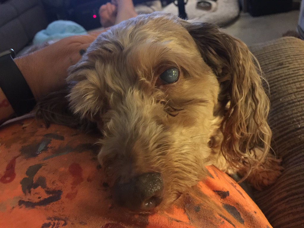
The cornea has the highest density of sensory nerve endings of any tissue in the body, which is why even a single misplaced lash causes immediate and intense awareness. In entropion, the entire lash line contacts the cornea with every blink - thousands of times per day.
The damage follows a predictable sequence. Chronic friction strips away the corneal epithelium, producing superficial punctate keratopathy visible as a constellation of tiny staining dots on slit-lamp examination. Clinical studies found corneal keratopathy in more than 60% of entropion patients, and dry eye syndrome in more than 70% - the two conditions reinforce each other, since a damaged epithelium cannot maintain a stable tear film and a poor tear film accelerates abrasion. Left longer, focal abrasions deepen into frank corneal ulcers, which are open portals for bacterial infection. A corneal ulcer that becomes infected can scar the visual axis permanently, causing vision loss that no subsequent surgery can fully reverse.
How a Surgeon Evaluates Entropion Before Choosing an Operation
The examination does not just confirm entropion - it determines which operation is appropriate. A surgeon who skips the mechanical tests and proceeds to a preferred procedure is guessing at what to fix.
- External inspection. The surgeon observes lid position at rest, in downgaze, and during active blinking. Involutional entropion often worsens dramatically in downgaze as the retractors lose the battle against the orbicularis. Visible lid scarring immediately raises the possibility of cicatricial disease.
- Snap-back test. The surgeon pulls the lower lid margin downward, releases it, and watches how quickly it returns. A lid with good tension snaps back at once. A lid that drifts back slowly - taking more than a second or two, or requiring a blink to return - indicates pathological horizontal laxity and identifies a patient who needs a structural tightening procedure.
- Distraction test. The lid margin is drawn outward from the globe and the displacement is observed. Normal movement is only a few millimeters. Significant laxity allows the margin to be pulled a centimeter or more away - a finding that makes the diagnosis of involutional entropion and the need for lid shortening essentially certain.
- Orbicularis override check. The patient gently closes the eye while the surgeon observes whether the preseptal orbicularis rolls over the pretarsal orbicularis. This confirms the third mechanical failure of involutional entropion and informs whether everting sutures need to be part of the repair.
- Slit-lamp examination with fluorescein staining. Dye applied to the cornea and viewed under cobalt blue light illuminates damaged epithelial cells. The distribution and density of staining shows how much corneal damage has already occurred and informs the urgency of repair.
Bridging the Gap: Temporary Relief Before Surgery
Surgery is the definitive answer, but it cannot always happen immediately. Temporary measures reduce corneal damage while waiting for an operative slot or while managing active inflammation.
- Lid-everting tape - adhesive strips applied to the lower lid skin pull the margin outward and hold it in a corrected position; requires daily reapplication and irritates the skin over time
- Lubricating drops and ointment - preservative-free drops during the day and a viscous ointment at night reduce friction between lashes and cornea without correcting position
- Bandage contact lenses - a soft lens placed on the cornea creates a physical barrier against lash contact; more effective than drops alone for significant keratopathy but carries infection risk with prolonged wear
- Botulinum toxin injection - injected into the lower orbicularis oculi, it weakens the muscle's ability to fold the lid inward; published data show an average correction duration of roughly 12 and a half weeks, ranging from 8 to 16 weeks depending on the individual, though studies vary and your surgeon can give you a realistic expectation based on your specific situation
Botulinum toxin does not interfere with or compromise the results of subsequent surgical repair, making it a safe bridge rather than a commitment to a particular path. It is not a substitute for surgery: the muscle recovers fully and the underlying structural failures are entirely unchanged when the effect wears off.
Surgery for Involutional and Spastic Entropion
The Quickert Suture: Effective for Mild Cases, Incomplete Alone
The Quickert everting suture procedure can be performed in a clinic room in roughly 15 minutes under local anesthesia. Three or four absorbable sutures are placed through the full thickness of the lower lid to re-establish the relationship between the retractors and the anterior lamella, rotating the margin outward. Relief is immediate. The procedure is appropriate for mild or intermittent entropion and for patients who cannot tolerate a longer operation.
The problem is durability. Quickert sutures do not address horizontal lid laxity. In patients with measurable laxity - the majority of those with involutional entropion - the underlying looseness reasserts itself and the lid folds inward again over months.
Lateral Tarsal Strip: Correcting the Structural Cause
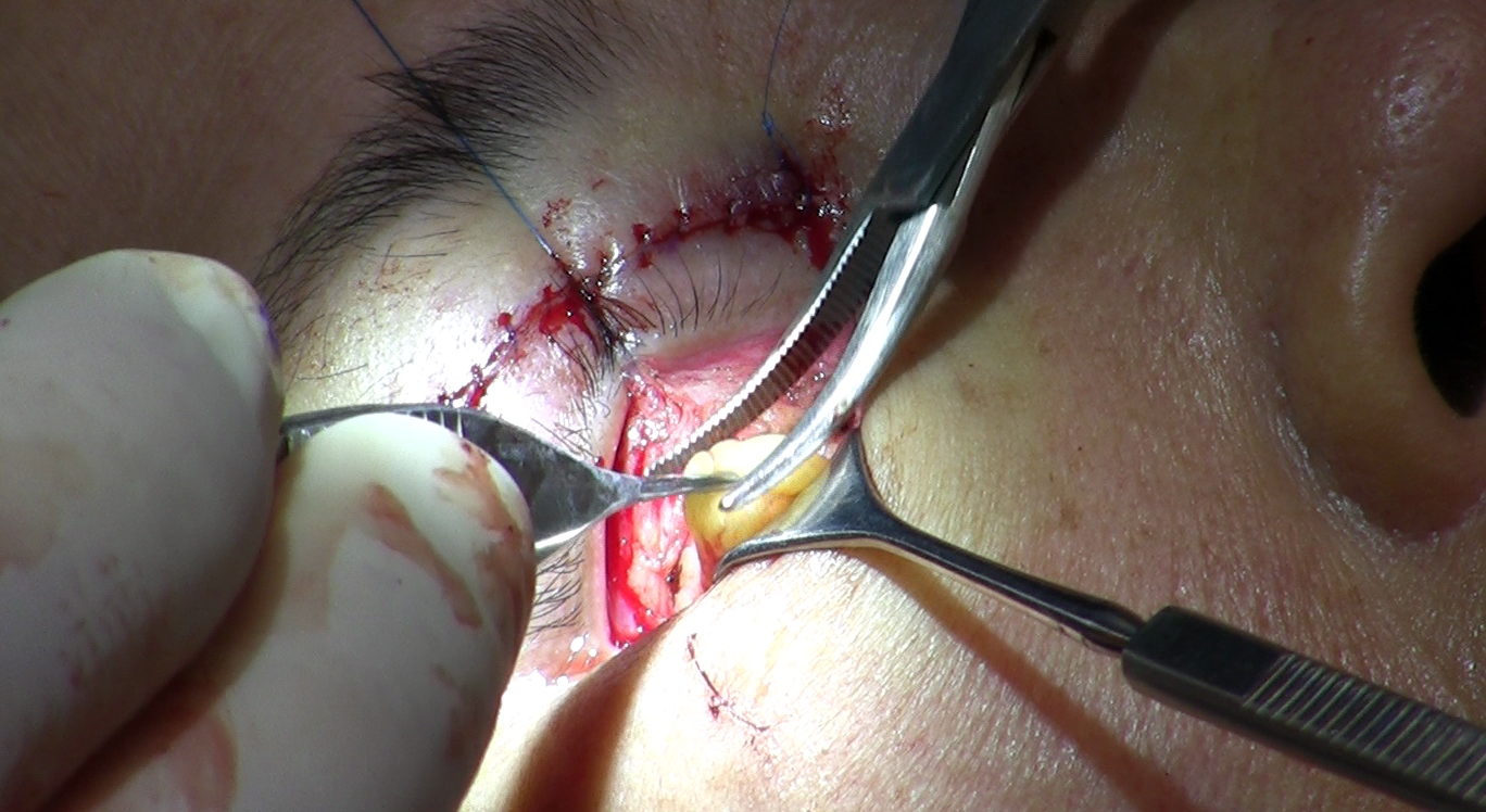
The lateral tarsal strip (LTS) procedure requires a small incision at the outer corner of the eye. The surgeon separates a strip of the tarsal plate - the rigid structural core of the lid - removes excess length from it, and anchors the shortened strip to the periosteum of the orbital rim. The lid is physically tightened and repositioned rather than held by a suture. Published studies generally report high success rates for LTS, with most series finding anatomical correction in over 90% of cases. Even so, published recurrence data for LTS alone vary by study and follow-up period, with some series reporting rates in the range of 10-20%, because horizontal laxity is only one of the three mechanical failures.
Randomized controlled data consistently support combining LTS with everting sutures: studies comparing different surgical approaches have found that combined procedures produce lower recurrence than either LTS or everting sutures performed alone. This is why experienced oculoplastic surgeons typically perform both in the same session: LTS corrects horizontal laxity and repositions the lid; everting sutures address the orbicularis override. Together they cover all three mechanical failures of involutional entropion.
Three separate mechanisms fail simultaneously in involutional entropion - horizontal laxity, retractor weakness, and orbicularis override - and a repair that addresses only one or two of them is a repair designed to recur.
Surgery for Cicatricial Entropion: A Different Operation Entirely
Cicatricial entropion requires tissue replacement, not tissue repositioning. The posterior lamella has been foreshortened by scar tissue. Tightening procedures move tissue around; they cannot restore tissue replaced by fibrosis. An LTS on a cicatricial lid would tighten an already contracted structure and worsen the inversion.
Anterior Lamellar Recession
In milder cicatricial cases, the surgeon separates the anterior lamella (skin and orbicularis muscle) from the posterior lamella and allows it to recess, releasing the pull between the two layers. This works when scarring is mild and the posterior lamella retains some elasticity. More advanced disease requires a different approach.
Mucous Membrane Grafting
Moderate to severe cases require a graft of tissue that matches the posterior lamella - moist, non-keratinizing mucosa. The two most common donor sites are the inner cheek (buccal mucosa) and the hard palate mucosa. The inner cheek provides a soft, supple graft suitable for most cases. Hard palate mucosa is firmer and better tolerated when structural support along the posterior lid surface is needed, particularly if the tarsus has been significantly damaged. The scarred posterior lamella is incised, the contracted tissue is released, and the graft fills the gap.
Recovery from cicatricial repair is longer than from involutional repair - often six weeks or more - because both the eyelid and the donor site must heal simultaneously. When the underlying disease, particularly Stevens-Johnson syndrome, remains active, new scarring can develop and staged or repeat procedures may be necessary.
Recovery: What Changes Immediately and What Takes Weeks
Most patients with involutional entropion notice relief within hours of the procedure, once the local anesthetic recedes. The foreign-body sensation, the constant awareness of something dragging across the eye, and the reflex tearing all disappear because lashes no longer contact the cornea from the moment the lid is repositioned. Full soft-tissue healing takes two to four weeks, with bruising and swelling around the outer corner of the eye fading across that period.
Watch for these signs during recovery:
- Return of the rubbing sensation - if the lid begins turning inward again within the first six months, it likely indicates recurrence; revision is feasible and worth discussing with the surgeon
- Increasing redness, discharge, or pain at the incision site - uncommon, but any worsening rather than improving wound appearance warrants prompt contact with the surgeon
- Persistent corneal symptoms beyond a few days - the epithelium needs time to regenerate; lubricating drops remain useful in the first weeks even after lid position is corrected
- Difficulty fully closing the eye - slight overcorrection sometimes occurs; this typically resolves as swelling subsides over the first two weeks
Entropion and Trichiasis: Where the Diagnosis Splits
Trichiasis - misdirected lashes from a normal lid - and entropion produce an identical chief complaint: lashes touching the cornea. The distinction is visible on examination. In entropion, the lid margin is rotated inward and the mechanical tests are abnormal. In trichiasis, the surgeon observes that the lid margin sits where it should, with no inward rotation and normal snap-back and distraction results, but specific lashes emerge at an angle directed toward the globe.
The treatments share no overlap. Trichiasis involving a small number of lashes is treated with electrolysis or cryotherapy directed at individual follicles to permanently destroy the misdirected root. Entropion is treated with the structural procedures described above. Performing cryotherapy on a truly entropic lid removes a few lashes while leaving the mechanical cause entirely in place. Performing LTS on a patient with purely trichiatic lashes tightens a structurally normal lid for no benefit.
Both conditions can coexist. A patient with long-standing involutional entropion may have individual lashes emerging at an abnormal angle from follicles altered by years of lid position change. The standard approach is to repair the entropion first and then reassess remaining lash misdirection once the lid sits in its corrected position - some apparent trichiasis resolves when the margin realigns, and any that persists can be targeted with follicle-specific treatment at the same session or shortly after.
Frequently Asked Questions
Can entropion go away on its own without treatment?
Congenital entropion in infants sometimes resolves in the first year as facial structure develops - this is the one form where watchful waiting is reasonable. In adults, involutional and cicatricial entropion do not self-correct; the mechanical failures that cause them do not reverse with time, and corneal damage accumulates throughout any delay. Spastic entropion may calm when the triggering inflammation is treated, but the underlying laxity it exposed generally requires surgical repair to prevent recurrence.
Why does my surgeon want to do two procedures rather than just one?
If you have involutional entropion with measurable horizontal laxity, everting sutures alone leave the laxity untreated - the most common cause of recurrence. The best available randomized trial data consistently show that combining lateral tarsal strip with everting sutures produces meaningfully lower recurrence than either procedure performed alone. Both procedures happen in the same operative session; the additional surgical time is small relative to the durability benefit.
How long does it take for the cornea to heal after entropion is corrected?
The corneal epithelium regenerates relatively quickly and most surface healing occurs within one to two weeks, aided by lubricating drops. Deeper corneal scarring, if it developed before surgery, does not reverse when the lid is corrected - the scar is structural and permanent. This is why early treatment matters: surface keratopathy heals, but established stromal scars do not.
What does recurrence actually look like, and is it treatable?
Recurrence typically presents as the return of the same foreign-body sensation and tearing the patient had before surgery - usually noticeable within the first six months rather than years later. The lid margin will be visibly rotating inward again on examination. Revision surgery is feasible when recurrence occurs; the surgeon may choose a different technique, a more extensive tightening, or add a procedure that was not included the first time. Early reporting of symptoms gives the most options.
My doctor mentioned I have both entropion and trichiasis - do I need two separate procedures?
Not necessarily in two separate visits. The standard approach is to repair the entropion first, then reassess individual lash position once the lid sits in its correct location. Some apparent trichiasis resolves when the lid margin realigns. Any lashes that remain misdirected can be targeted with electrolysis or cryotherapy - sometimes during the same session, sometimes as a brief follow-up procedure after healing is confirmed.
General information only, not medical advice. Individual anatomy and healing vary. See the disclaimer.