A drooping upper eyelid is one of the most precisely correctable structural problems in facial surgery - but only when the surgeon identifies which part of the lifting mechanism has actually failed. Three fundamentally different operations address ptosis, each targeting a different anatomical structure. Which one a patient receives is not a matter of surgical preference; it follows directly from objective measurements taken in the exam room, numbers that define where the system broke down and how much capacity remains to restore it.
Ptosis and Blepharoplasty: The Same Eyelid, Two Different Problems
Excess skin resting on the lashes and a lid margin that sits too low look similar from the outside. They are not the same problem, and treating one when the other is present will not help.
Blepharoplasty removes redundant skin and fat from the upper lid - a tissue-volume correction. Ptosis repair addresses a mechanical failure: the apparatus that lifts the lid has either disconnected from its anchor point, weakened past the point of effective contraction, or lacks meaningful function from birth. Removing skin does nothing for lid height when the real problem is a detached tendon. A surgeon who operates on the muscle when only tissue needs removing can introduce a lid-position complication where none existed.
Some patients genuinely need both procedures at the same sitting - but only when each problem is confirmed independently, not assumed because the eye looks tired. The insurance implications follow the same logic. Ptosis repair can qualify as a functional procedure when it causes measurable visual obstruction; cosmetic blepharoplasty does not. A formal visual field test performed with the lid in its natural, drooping position - documenting significant loss in the upper field of vision - is typically required to support a functional claim. Check current documentation requirements directly with the insurer and surgeon, as thresholds vary by plan.
What Surgeons Measure Before Deciding
Every oculoplastic evaluation for ptosis centers on two core measurements. Together they define the severity of the droop and the remaining capacity of the lifting muscle - the two variables that determine which operation is offered.
MRD1: Quantifying the Droop
MRD1, or marginal reflex distance 1, is the gap between the corneal light reflex and the upper lid margin when the patient looks straight ahead into a light source. Normal falls at roughly 4-5 mm. A shortfall of 2 mm from the expected upper range classifies as mild ptosis, 3 mm as moderate, and 4 mm or more as severe. The number gives the surgeon a baseline and a target - it reveals nothing about cause.
Levator Function: The Decision Point
Levator function is measured as the total lid excursion from full downgaze to full upgaze while the examiner's thumb firmly holds the brow down. That brow pressure is not incidental - it blocks the frontalis muscle from secretly augmenting the movement. Without it, a patient with near-zero levator capacity can produce a misleadingly passable number through forehead compensation alone. The results fall into clear grades:
- Poor - less than 4 mm of excursion
- Fair - 5 to 9 mm
- Good - 9 to 11 mm
- Excellent - 12 mm or above
- Normal - exceeds 15 mm
This single number is the decision point. Good or excellent levator function opens the door to techniques that work by adjusting the levator mechanism. Poor function - under 4 mm - means there is not enough working muscle to adjust or stimulate. A completely different mechanical solution is required. Additional lower-lid assessments, including snap-back and distraction tests that check for laxity, round out the pre-operative picture when lower-lid work or orbital fat repositioning is also being considered.
| Technique | Levator Function Needed | Ptosis Severity | Incision Site | External Scar |
|---|---|---|---|---|
| Levator Advancement | Fair to excellent (5 mm+) | Mild to severe | Eyelid crease | Yes, hidden in crease |
| MMCR | Good to excellent (10 mm+) | Mild to moderate | Inside the lid | None |
| Frontalis Sling | Poor (under 4 mm) | Moderate to severe | Lid, brow, forehead | Small, multiple sites |
The Phenylephrine Test: What It Reveals and Why It Underestimates
Muller's muscle is a thin band of smooth muscle in the upper lid, controlled by the sympathetic nervous system independently of the voluntary levator. It contributes roughly 1 to 2 mm of elevation at full contraction - a figure consistent across standard anatomy references. Because it is anatomically and neurologically separate from the striated levator muscle, it can be specifically targeted - and specifically tested.
Before offering MMCR, surgeons instill phenylephrine drops into the affected eye. The standard protocol applies either 2.5% or 10% concentration; the specific number of drops and timing between instillations vary by practice and published protocol, so confirm the details with your surgeon. Because phenylephrine stimulates only sympathetically innervated tissue, any lid rise it produces measures Muller's isolated contribution. The levator is untouched. If the lid reaches an acceptable height, the patient is a candidate for MMCR.
The test has a known and clinically important limitation: it consistently underestimates the surgical outcome. Drops produce an average lid rise substantially smaller than what the operation achieves - published studies vary on how large that gap is, but the directional bias is well documented. A borderline positive test in the office reliably predicts a good surgical result. Surgeons who dismiss candidates because the office response seemed modest may be turning away patients who would have done well.
Levator Advancement: Reattaching the Tendon Through the Eyelid Crease
Levator advancement is the most common ptosis repair technique. It is appropriate when the levator muscle retains fair to excellent function but has lost its connection to the lid - the classic picture of aponeurotic ptosis, where the levator aponeurosis stretches or separates from the tarsal plate over years and migrates upward, leaving the lid without traction from above.
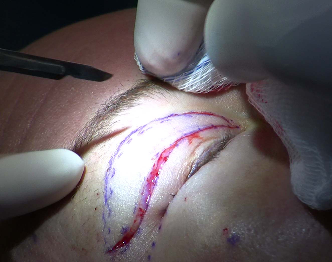
The operation proceeds through the natural eyelid crease, which hides the incision afterward. The sequence:
- Local anesthetic is injected into the upper lid while the patient remains awake.
- The surgeon opens the crease incision and dissects through orbicularis muscle to reach the orbital septum.
- The septum is opened; the preaponeurotic fat pad is identified - a key anatomical landmark confirming the correct surgical plane.
- The levator aponeurosis is located. In aponeurotic ptosis it will be thinned, stretched, or visibly separated from the tarsal surface.
- The aponeurosis is sutured back to the upper third of the tarsal plate using a double-armed absorbable suture, placed but not finally tied.
- The patient sits upright and looks straight ahead. The surgeon assesses lid height and contour before committing.
- Sutures are adjusted based on what the upright, awake patient shows - the lid can be nudged higher or eased lower before final closure.
Step six is what separates this operation from almost every other reconstructive procedure. Lid height under local anesthesia with the patient sitting is a direct, real-time measurement of the functional outcome. A result that looks symmetric on the operating table with the patient flat can look quite different when the patient is vertical and the lid is working against gravity. The sitting adjustment is not a contingency measure - it is a deliberate part of how the technique achieves accuracy.
Muller's Muscle Conjunctival Resection: The Inside Approach
MMCR reaches the lid from behind, through the conjunctival surface, with the lid everted to expose its inner face. No scalpel touches external skin. No visible scar forms on the outside of the eyelid.
The right candidate for MMCR has a specific profile:
- Mild to moderate ptosis, generally in the 2-3 mm range
- Good to excellent levator function - typically 10 mm or above
- A positive or borderline-positive phenylephrine test
- No preference for or requirement of simultaneous crease reformation
When those criteria are met, MMCR tends to outperform the external approach on objective metrics. In published comparative studies, MMCR generally achieves higher surgical success rates than external levator advancement; reported figures vary considerably by study design and patient selection, so ask your surgeon about current evidence for your specific presentation rather than relying on any single published figure.
The operation itself is efficient. After the lid is everted, a Putterman clamp is applied across the conjunctiva and Muller's muscle, positioned after marking resection points nasal and temporal to the steepest midpoint of the lid. The clamped tissue is excised and the cut edge closed. Because there is no skin incision, the crease line shows less bruising and swelling during recovery. The trade-off is specificity: MMCR corrects only a modest degree of ptosis and works only when Muller's muscle retains meaningful capacity. Larger ptosis or reduced levator function disqualifies a patient regardless of the desire to avoid a visible scar.
The Frontalis Sling: Bypassing the Levator Entirely
When levator function falls below 4 mm, there is simply not enough working muscle to adjust or stimulate. Levator advancement would find nothing functional to advance. MMCR would produce no meaningful elevation. These patients need a mechanism that bypasses the levator system and provides an entirely different source of lid-opening force.
A frontalis sling physically links the upper lid to the frontalis muscle of the forehead. When the patient lifts their brow, the sling transmits that movement downward and raises the lid. The connection is typically made with a strip of fascia lata - connective tissue taken from the outer thigh - or with a synthetic material in young children or cases where harvesting sufficient autologous tissue is not practical. Small incisions at the lid margin, brow, and forehead allow the material to be tunneled into position.
The resulting lid movement feels and looks different from normal. The lid and brow move together; raising the brow opens the eye, and looking downward may lower the lid somewhat. Incomplete closure during sleep - nocturnal lagophthalmos - is common and typically managed with lubricating ointment on an ongoing basis. These are not surgical complications; they are the expected functional profile of a lid that opens by brow elevation rather than levator contraction. Frontalis sling is the standard approach for congenital ptosis with poor levator function, frequently performed in children whose visual development depends on adequate pupil exposure.
Recovery: The Swelling Arc and What to Watch For
Ptosis repair runs roughly 45 to 90 minutes per eye as an outpatient procedure. Most patients go home the same day. The final lid position takes weeks to settle and should not be evaluated too early.
- First two to three days: Swelling and bruising peak. The lid may look higher than expected - swelling-related apparent overcorrection at this stage is normal.
- Dry eye risk: A higher lid exposes more corneal surface. Lubricating drops are nearly universal in the early weeks; symptoms are usually transient but can persist several months in patients with pre-existing tear film issues.
- Four to six weeks: Swelling continues to resolve and the lid settles toward its final position. Most surgeons defer formal outcome assessment until at least six to eight weeks post-operatively.
- Overcorrection: The lid sits too high, exposing a wider band of white below the iris than normal. Most cases resolve with continued healing; persistent overcorrection may need a small revision.
- Undercorrection: The lid remains lower than the target. More common than overcorrection, and typically addressed with a secondary procedure after full healing.
Durability and the Risk of Recurrence
Ptosis repair is not uniformly permanent. Research tracking outcomes over a median follow-up of roughly three years documents recurrence in approximately 23% of operated eyes - and the rate varies considerably based on the underlying cause.
Aponeurotic ptosis - where the levator tendon detaches from the tarsal plate over time - recurs at substantially lower rates than congenital or myogenic ptosis, where the muscle itself is the primary site of failure.
This distinction matters for setting expectations. A patient whose lid drooped gradually in their fifties due to age-related tendon changes is a better candidate for durable long-term correction than someone whose ptosis stems from a muscle that never developed normally. The levator mechanism in aponeurotic cases is structurally intact; reattaching the tendon restores a working system. In congenital or myogenic cases, the underlying deficit persists.
One recurrence risk that patient-facing guides almost never mention: intraocular surgery performed after ptosis repair significantly raises the chance of reoperation. Patients who later undergo cataract extraction face a meaningfully higher reoperation rate than those who do not, an effect documented at five and ten years of follow-up. Surgical manipulation of the eye during cataract surgery is thought to stress the repaired levator attachment. Patients who anticipate needing cataract surgery in the near future should discuss the sequencing explicitly with both surgeons - when each procedure happens relative to the other can affect how long the ptosis result holds.
Frequently Asked Questions
If I have ptosis in one eye, can fixing it cause the other eye to droop?
Yes, and this is one of the most important pre-operative conversations to have. The brain sends equal neural drive to both levator muscles simultaneously - a principle called Hering's law of equal innervation. When one eye droops, the brain increases its signal to lift it, and that elevated drive raises the fellow eye as well, masking whatever droop it would otherwise show. After the ptotic eye is repaired, the extra neural demand disappears, and the previously masked droop in the other eye can become visible for the first time. Surgeons routinely test for this before surgery by manually lifting the ptotic lid and watching what the fellow eye does: if it falls, the patient should understand that a second procedure on the other side may follow the first. Knowing this in advance eliminates the most common source of post-operative surprise in unilateral ptosis repair.
When does a drooping eyelid signal a neurological problem rather than a mechanical one?
Three neurological conditions cause ptosis through pathway failure rather than structural wear, and each requires evaluation before any surgical plan is formed. Third nerve palsy typically presents with ptosis combined with limitation of eye movement and often a dilated, poorly reactive pupil - that combination points to the oculomotor nerve and warrants urgent imaging to exclude a compressive cause. Horner's syndrome produces mild ptosis alongside a smaller pupil on the same side, without significant reduction in levator excursion; the lesion can sit anywhere along the sympathetic chain from the brainstem to the orbit, and the underlying cause ranges from benign to serious. Myasthenia gravis causes fatigable ptosis that worsens over the course of a day and after sustained upgaze, can alternate between sides, and is often accompanied by other muscle weakness. A new-onset drooping lid with any accompanying pupil asymmetry, double vision, or fatigue-related variability is not a cosmetic finding until neurological causes are excluded - surgeons screen for all three as part of the standard pre-operative assessment.
How is ptosis in a child different from ptosis in an adult?
The stakes and the timeline are fundamentally different. In adults, a drooping lid causes cosmetic concern and may obstruct the upper visual field, but the visual system is fully mature - delayed repair carries no developmental risk. In children, a lid that covers the visual axis or induces a habitual chin-up head posture to see can deprive the eye of the patterned visual input it needs during the critical window for visual development, leading to amblyopia - a reduction in best-corrected vision that glasses alone cannot fix. The urgency of repair scales with how much of the visual axis is blocked and whether amblyopia is already developing; some cases require intervention in infancy. Children with congenital ptosis and poor levator function receive frontalis slings rather than levator advancement, and most display lid lag on downgaze - the affected lid does not drop fully as the eye moves down, because the fibrotic levator muscle lacks normal elasticity. Follow-up typically extends through the school years, and reoperation as the face grows is common.
Does ptosis repair leave a visible scar?
It depends on the approach. Levator advancement places an incision in the natural eyelid crease, where healed scar tissue is typically invisible once fully mature. MMCR leaves no external incision - the procedure is done entirely from inside the everted lid. A frontalis sling requires small incisions at the lid margin, brow, and forehead; these fade but are more numerous than the other two approaches.
Are there non-surgical options for a drooping eyelid?
One exists for patients who are not surgical candidates or who are not yet ready to commit to an operation: a ptosis crutch, which is a small wire or acrylic attachment fitted to an eyeglass frame that physically props the upper lid open. It requires glasses to be worn at all times the correction is needed, must be periodically adjusted as the lid position or frame changes, and corrects nothing structurally - the lid returns to its resting position whenever the glasses come off. Its practical role is as a bridge for elderly patients whose medical condition makes anesthesia high risk, or as a temporary measure while a surgical decision is being made. No topical medication or injection reliably raises lid height the way surgery does. Onabotulinumtoxin A is sometimes mentioned in this context, but its established role in eyelid position runs the other direction: it is a recognized cause of temporary ptosis when it migrates from brow or glabellar injection sites, not a corrective treatment for it.
General information only, not medical advice. Individual anatomy and healing vary. See the disclaimer.