When a surgeon describes lower eyelid surgery, the conversation usually focuses on fat pockets, tear troughs, or loose skin. What rarely gets explained - and what shapes almost every downstream outcome - is where the first incision actually goes. Lower blepharoplasty can be performed through the inner mucosal surface of the eyelid, leaving the outer skin completely unmarked, or through the outer skin along the lash line, producing a scar that takes months to mature. That single anatomical decision determines whether a key structural layer called the orbital septum stays intact, whether any skin can realistically be removed at all, and how likely the lower eyelid is to pull downward after surgery. Most patients arrive at consultations without knowing the choice exists.
The Incision Route as a Separate Decision
Surgeons performing lower blepharoplasty are always making two distinct choices: what to do with the fat compartments, and how to get there. The fat plan - whether to trim excess volume, reposition fat over the orbital rim to fill a tear trough, or leave it alone - is one question. The incision route is a separate, upstream question, and the answer constrains what the fat plan can even accomplish.
A surgeon who chooses the transconjunctival route gains direct access to all three lower eyelid fat compartments from the inside, avoids cutting the skin entirely, and preserves a fibrous structural layer whose disruption is the primary source of post-operative eyelid retraction. A surgeon who chooses the transcutaneous route gains the ability to excise actual skin but accepts the anatomical consequences of opening that same layer. Both are legitimate tools in the right hands and the right patient. The problem is that patients are rarely told which is planned - or why - until they specifically ask.
The Transconjunctival Approach: Where the Cut Goes and Why No Scar Results
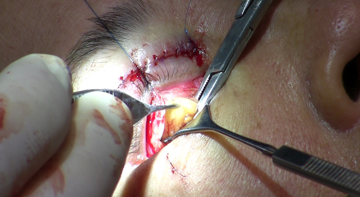
The transconjunctival incision is placed entirely on the conjunctiva - the moist mucosal lining on the inner surface of the lower eyelid. The outer eyelid skin is never cut. From the patient's perspective, there is no visible entry point at all once the eyelid is closed.
Once through the conjunctiva, the surgeon has a direct line of sight to the three orbital fat compartments: the medial, central, and lateral pockets. Each can be individually accessed, trimmed for volume reduction, or mobilized and repositioned downward over the orbital rim to fill a tear trough depression. That repositioning maneuver involves cutting the arcus marginalis - the fibrous periosteal attachment of the septum at the orbital rim - and can be performed entirely through the conjunctival incision without leaving any mark on the skin surface.
When the procedure is complete, the conjunctival incision is typically closed with absorbable sutures such as plain gut that dissolve on their own, or in some cases the wound is left to self-seal. No suture removal appointment is required. The absence of a skin incision is not merely cosmetic - it reflects a fundamentally different anatomical path that leaves the structural layer separating the fat from the skin completely undisturbed.
The Transcutaneous Approach: Subciliary, Subtarsal, and What the Scar Actually Does Over Time
Transcutaneous lower blepharoplasty cuts through the outer skin to reach the underlying fat and, when necessary, to remove a strip of redundant skin. Surgeons have two main placement options for this external incision, and they are not equivalent in terms of scar placement or visibility.
Subciliary Incision
The subciliary approach places the cut just below the lash margin, running closely along the lower lash line. In the early post-operative weeks, the scar appears as a pink or reddish line tracking the lid margin. Over several months it flattens and pales. In patients with mature skin texture - where natural lines and pores provide camouflage - the final faded scar can be difficult to spot. In younger patients with smooth, unlined skin, the scar remains more conspicuous during the maturation period, which can extend to a year.
Subtarsal Incision
The subtarsal approach places the cut several millimetres below the lashes, within a natural skin crease. Some surgeons favor this position precisely because the crease provides a topographic hiding place for the healing wound. The scar sits lower on the lid, which changes the geometry of skin excision. Both transcutaneous variants share the same critical deep consequence: to reach the fat compartments, the surgeon typically opens the orbital septum, with the degree of disruption depending on the specific technique used.
The Orbital Septum and Middle Lamella: The Anatomy That Explains the Risk Gap
The orbital septum is a fibrous sheet running from the orbital rim and attaching to the lower eyelid retractors near the inferior edge of the tarsus. It acts as the boundary between the orbital fat compartments behind it and the anterior lamella - the skin-and-muscle layer of the eyelid's outer surface. When intact, the anterior lamella maintains its vertical length and the lower lid holds its normal position.
Any transcutaneous approach that opens this septum allows it to heal with scar tissue. That scar tissue contracts. Contracture shortens the anterior lamella vertically, and a shortened anterior lamella can no longer hold the eyelid margin in its correct position. The result is lower lid retraction - the lid margin pulls downward, exposing sclera below the iris - or, in more severe cases, ectropion, where the lid actually turns outward.
Published surgical literature, including the StatPearls review available on NCBI, documents substantially higher lower eyelid retraction rates with the subciliary transcutaneous approach compared to the transconjunctival route - readers should consult those sources directly for the specific figures cited in current literature. The transconjunctival route, which leaves the orbital septum entirely intact, avoids triggering this contracture mechanism altogether.
The orbicularis oculi muscle adds a second layer to this risk. It wraps around the periorbital area and provides active muscular support for the lower lid margin. Transcutaneous flap elevation that splits or denervates this muscle can weaken that active support, reducing the lid's ability to resist the downward pull of post-operative scar forces.
What Each Approach Can and Cannot Treat
The right approach depends on what the patient's anatomy actually requires. These two routes do not treat the same problems equally, and conflating them leads to both under-treatment and avoidable complications.
The Transconjunctival Approach Addresses
- Herniated orbital fat causing under-eye puffiness or visible bags
- Tear trough hollowness, through fat repositioning via arcus marginalis release
- Mild to moderate skin quality issues, when combined with CO2 laser resurfacing or a skin pinch
- Fat compartment volume asymmetry between the two sides
The Transconjunctival Approach Cannot Address Alone
- Significant skin redundancy - loose skin will remain loose because no skin is excised
- Pronounced orbicularis muscle hypertrophy or festoons - there is no direct access to the muscle from the conjunctival side
- Severe skin wrinkling that exceeds what laser resurfacing can correct
The Transcutaneous Approach Addresses
- Substantial skin excess requiring formal excision - the defining reason to accept the higher retraction risk
- Orbicularis muscle bands or rolls that need direct surgical reduction
- Combined skin and fat problems managed within a single operative field
- Patients with good lid tone and no high-risk anatomical factors who can tolerate septum disruption without significant retraction risk
The practical sorting principle: if the primary concern is fat herniation in a patient with good skin quality and reasonable lid tone, the transconjunctival route almost always wins on the risk calculation. If loose, excess skin is the dominant complaint and conservative alternatives cannot fully address it, the transcutaneous route - with its ability to excise skin - may be the only path to a complete correction.
Who Belongs in Which Column
Several clinical factors push patients toward one approach or the other. A thorough preoperative assessment will examine all of them before an incision route is settled.
Factors Favoring the Transconjunctival Route
- Negative vector anatomy: When the globe protrudes forward of the malar eminence, the cheek provides less mechanical support to the lid. Septum disruption in this anatomy raises retraction risk to a degree that most surgeons consider unacceptable when the transconjunctival route can achieve the surgical goals.
- Pre-existing dry eye syndrome: Even minor post-operative lid retraction produces lagophthalmos - incomplete eyelid closure - which exposes the cornea and worsens dry eye symptoms dramatically. Transconjunctival surgery carries significantly less lagophthalmos risk and is the strongly preferred route for patients with a documented dry eye diagnosis.
- Primary fat complaint with acceptable skin quality: Patients whose main concern is puffiness without meaningful skin laxity are ideal transconjunctival candidates, often with laser or skin pinch added for surface improvement.
- Younger patients: Younger skin has good elasticity and little redundancy. Fat-only correction is typically sufficient, and avoiding a lash-line scar during the decades when it would be most visible is a meaningful benefit.
Factors Favoring the Transcutaneous Route
- Significant skin redundancy: When the primary correction requires removing skin, a transcutaneous approach is typically the only way to accomplish it.
- Strong lid tone on preoperative testing: Good canthal tendon integrity means the lid has the mechanical reserve to resist retraction forces after septum disruption.
- Orbicularis hypertrophy or festoons: When the muscle layer itself needs direct surgical reduction, the transcutaneous flap provides the access the conjunctival route cannot offer.
Before either approach is finalized, a snap-back test and distraction test are performed on the lower lid to assess canthal laxity. Significant laxity - where the lid can be pulled a clinically meaningful distance from the globe - signals that a canthopexy or canthoplasty is needed at the same time as the blepharoplasty, regardless of which incision route is chosen. Operating on a lax lid without addressing the underlying laxity is one of the more preventable contributors to poor results.
The Skin Pinch Hybrid and CO2 Laser: Addressing Skin Without Giving Up Safety
For patients with primarily a fat problem but also mild to moderate skin laxity, two adjunct techniques allow surgeons to address the skin without crossing into the higher-risk territory of formal transcutaneous surgery.
Skin Pinch Blepharoplasty
In a skin pinch, the surgeon completes the full transconjunctival procedure first, then - without raising a skin-muscle flap and without cutting the orbital septum - uses fine forceps to pinch and excise a small ellipse of redundant skin just below the lash line. Because no deep dissection accompanies this skin removal, the middle lamella and septum remain undisturbed. The patient gets modest skin reduction with the structural protection of the transconjunctival approach intact. A skin pinch is not a replacement for formal skin excision in patients with marked redundancy - it handles mild excess, not the full range that a transcutaneous flap can address.
CO2 Laser Resurfacing as a Same-Session Adjunct
CO2 laser resurfacing of the lower eyelid skin can be performed in the same operative session as transconjunctival blepharoplasty. The laser treats fine lines, mild wrinkling, and pigment changes through controlled thermal remodeling rather than excision. It produces measurable improvement in skin texture and some degree of tightening without a scalpel ever crossing the skin surface. For patients with fat herniation and skin quality concerns that fall short of requiring excision, this combination targets both complaints while maintaining the septum-intact safety profile of the conjunctival incision.
The skin pinch and CO2 laser adjuncts exist precisely to give surgeons a middle path - addressing skin laxity and surface quality without triggering the scar contracture mechanism that makes formal transcutaneous surgery riskier than the transconjunctival baseline.
Recovery Compared Side by Side
| Factor | Transconjunctival | Transcutaneous (Subciliary) |
|---|---|---|
| External scar | None | Lash-line scar; matures over months |
| Suture removal needed | No - dissolving or self-sealing | Yes - typically at 5 to 7 days |
| Bruising timeline | Moderate; usually clears in 1-2 weeks | Often heavier; may last 2-3 weeks |
| Swelling resolution | Most resolves in 2-3 weeks; residual fades longer | Similar; scar thickening adds visible changes |
| Lower lid retraction risk | Low (septum left intact) | Substantially higher - see StatPearls/NCBI for cited rates |
| Skin removal capability | Limited (skin pinch only) | Yes - formal excision possible |
The early post-operative appearance of a subciliary scar moves through predictable stages. In the first two weeks it may appear pink, slightly raised, and more obvious than patients expect. By six to eight weeks it typically flattens and begins to pale. Full maturation - where the scar reaches its lightest, least visible final state - can take six months to a year. Patients who have not been briefed on this timeline often become alarmed at the four-week mark, when the scar is still in an active remodeling phase and looks worse than it will at twelve months.
With the transconjunctival approach, recovery is primarily about bruising and swelling rather than wound healing. Chemosis - swelling of the conjunctiva itself - can occur after the internal incision and typically resolves within a few weeks, usually managed with conservative measures such as lubricating drops and cold compresses. Patients frequently note that the lack of a visible wound or sutures makes the recovery feel less severe, even when the underlying swelling is comparable.
Questions to Ask Your Surgeon Before Lower Eyelid Surgery
Patients who ask specific, targeted questions before lower blepharoplasty leave the consultation with a clear picture of what is planned and why. These questions, asked in roughly this order, will reveal whether the surgeon has thought through your anatomy specifically rather than applying a default approach.
- Which incision route are you planning - transconjunctival or transcutaneous - and what specific finding in my anatomy drove that recommendation? The answer should name one route and cite specific observations: lid tone on testing, the degree of skin laxity, fat distribution across compartments, or a structural factor like facial vector anatomy.
- Will the orbital septum be disturbed during this procedure? If yes, ask what the surgeon does to minimize retraction risk and what the signs of retraction would look like if it occurred.
- Did you perform a snap-back and distraction test on my lower lid, and what did those tests show? If canthal laxity is present, ask whether a canthopexy or canthoplasty is included in the surgical plan.
- Do I have negative vector anatomy, and does that change which approach is safer for me? A surgeon who does not have a clear answer to this question is worth questioning further before committing to surgery.
- I have dry eye - how does that factor into your approach recommendation? If you have a diagnosed dry eye condition, this should be part of the pre-operative planning conversation explicitly, not an afterthought.
- If I have mild skin laxity but not severe laxity, am I a candidate for a skin pinch or CO2 laser combination rather than full transcutaneous surgery? Many patients who assume they need formal skin excision can be well-served by a hybrid approach that preserves the septum.
- Walk me through what my recovery will look like week by week, including what the scar or swelling will look like at four weeks and at three months. A surgeon who can describe this specifically has been through it enough times to have a reliable clinical picture of the outcome.
Frequently Asked Questions
Is transconjunctival blepharoplasty less effective than transcutaneous because it leaves the skin alone?
Not for the right patient. In patients whose primary concern is fat herniation or tear trough hollowness with reasonably good skin quality, transconjunctival surgery produces complete correction of those specific issues without touching the skin. The approach falls short only when significant skin redundancy is also present - in that case, it needs a skin pinch or laser adjunct to address surface concerns, or a formal skin excision becomes necessary.
Can a subciliary scar realistically become invisible after it heals?
In many patients with mature skin texture and good wound healing, the subciliary scar becomes very difficult to detect once fully matured - a process that typically takes six months to a year. In patients with smooth, unlined, or younger skin, the scar tends to remain more visible even after full maturation. Post-operative sun protection during the maturation period meaningfully affects the final result.
What is negative vector anatomy and how would I know if I have it?
Negative vector describes a facial structure where the globe of the eye sits forward of the cheekbone - the malar eminence. Looking at your profile in a mirror gives a rough indication: if the eye appears to protrude relative to the cheek, you may have this anatomy. A formal assessment requires direct clinical evaluation, because this structural feature changes the mechanical support available to the lower lid and raises retraction risk substantially with transcutaneous approaches.
If I have dry eye, can I still have lower eyelid surgery?
Dry eye is a relative contraindication to the transcutaneous approach, not an absolute barrier to lower eyelid surgery altogether. The transconjunctival route carries significantly less risk of post-operative lagophthalmos - incomplete eyelid closure - which is the mechanism that worsens dry eye after transcutaneous surgery. Patients with a dry eye diagnosis should disclose it during the consultation so the surgeon can factor it explicitly into the approach decision; a pre-operative ophthalmology evaluation is often appropriate.
Why would a surgeon recommend transcutaneous surgery if the retraction risk is higher?
Because for some patients - particularly those with significant skin excess, strong lid tone, and no high-risk anatomical factors - the transcutaneous approach is the only method that achieves a complete result. The goal is not to eliminate all risk but to match the risk profile to the patient's anatomy and what the surgery needs to accomplish. The problem arises when the approach is applied broadly without patient-specific assessment rather than reserved for cases where it is the right tool.
What is a canthopexy and when does it need to accompany blepharoplasty?
A canthopexy tightens the lateral canthal tendon - the fibrous attachment at the outer corner of the eye that anchors the lower lid in position - without detaching it. This is the less invasive of two related procedures. When laxity is more significant, a canthoplasty may be performed instead, which involves cutting, shortening, and reattaching the tendon to restore adequate support. When the snap-back or distraction test reveals laxity in this tendon, the lower eyelid lacks the structural support needed to resist downward scar forces after surgery. Performing blepharoplasty in that setting without addressing the laxity - regardless of which incision route is chosen - increases the risk of lid malposition meaningfully. Most surgeons perform the canthopexy or canthoplasty in the same operative session rather than staging it separately.
General information only, not medical advice. Individual anatomy and healing vary. See the disclaimer.