The American Academy of Ophthalmology describes double eyelid surgery as one of the most frequently performed cosmetic eyelid procedures in the world. The procedure addresses a single anatomical fact: roughly 40 to 60 percent of East Asian individuals are born without a supratarsal crease. Most patient guides explain this as 'different anatomy' and stop there. This article goes further - into the specific tissue layer that determines whether a crease forms, how surgeons measure and plan the result in millimeters before the operation begins, what each technique actually does to the eyelid structures, and the complications that most patient-facing pages ignore entirely.
Why Some Asian Eyelids Have No Crease
The crease forms - or fails to form - based on one anatomical variable: the height at which the orbital septum fuses with the levator aponeurosis.
The levator muscle elevates the lid. As it descends toward the lid margin, it flattens into a broad fibrous sheet called the levator aponeurosis. The orbital septum is a thin fibrous membrane that separates the bony orbit from the eyelid soft tissue. In eyelids that develop a natural crease, these two structures fuse 8 to 10 mm above the upper border of the tarsal plate. Distal fibers from the aponeurosis extend outward through the orbicularis muscle and insert on the underside of the skin. When the lid rises, those fibers pull the skin inward and create the visible fold.
In monolid Asian eyelids, the septum-levator junction sits lower - fewer than 8 mm above the tarsal border. That lower fusion point means the distal aponeurosis fibers do not reach the skin in the same way. More consequentially, it leaves the preaponeurotic fat pad without a firm barrier holding it up. The fat descends toward the lash line, filling the space between the lashes and the brow and obscuring what would otherwise be a visible lid platform.
This is not a minor variation. The descending fat pad is what gives the monolid its characteristic fullness. The prevalence of this anatomy varies by population: rates are around 17 percent in some Japanese cohorts and close to 59 percent in northern Chinese populations, with a clear north-to-south gradient across China that reflects regional genetic differences.
The Four Fat Compartments of the Asian Upper Eyelid
Upper eyelid fat is not a single mass. Asian lids contain four anatomically distinct fat compartments, and knowing which one does what matters for surgical planning and for understanding complications.
- Subcutaneous fat: just beneath the skin surface, thin in most lids
- Pretarsal fat: overlying the tarsal plate in the lower portion of the lid
- Preaponeurotic fat: the pad that sits directly in front of the levator aponeurosis, the one most discussed in standard blepharoplasty
- Submuscular fibroadipose tissue (SMFAT): the layer between the orbicularis muscle and the orbital septum
In Caucasian eyelids, the SMFAT layer is thin and has little surgical significance. In Asian eyelids, it is the main source of upper eyelid thickness - the tissue most responsible for the characteristic fullness. When a surgeon fails to recognize and adequately address SMFAT during an incisional procedure, the bulk remains beneath the new crease and pushes the pretarsal skin outward. That is the direct mechanism behind sausage eyelid, described in detail later.
Fat preservation matters in the opposite direction too. Aggressive removal of the preaponeurotic fat pad hollows the upper eyelid and produces a sunken, skeletal appearance that reads as unnaturally Westernized. This is one of the most common reasons patients request revision. Asian double eyelid surgery preserves the preaponeurotic fat or reduces it only modestly.
Designing the Crease Before Surgery
Crease Height
Crease height is measured from the upper lash line - the gray line - to the planned crease. Most surgeons set it at 3 to 5.5 mm for male patients and 5 to 7.5 mm for female patients when the goal is a natural-looking Asian crease. Heights of 8 to 10 mm produce a 'Western' appearance - bolder, more conspicuous, and a frequent source of revision requests when patients feel the result does not look proportionate to their face.
This is not about ethnic categories as such. It is about structural proportion. A crease set at 9 mm on an eyelid whose natural lid platform is 6 mm wide looks incongruous. Patients who ask for 'a bigger crease' often mean they want a more visible crease, not a higher one. Clarifying that distinction before surgery matters.
Crease Shape
Two shapes are used in practice. The tapered crease starts narrow at the inner corner and widens toward the outer corner, following the natural slope of Asian lid anatomy. The parallel crease maintains a consistent height from inner to outer corner, producing a bolder, more defined look. When a natural result is the goal, the tapered crease is more often chosen. The parallel crease is not wrong - but it is more conspicuous, and patients should see both options marked on their own lids before deciding.

The Suture (Non-Incisional) Technique
The suture technique creates the crease without a full skin incision. The surgeon makes three to five small puncture holes along the planned crease line. Through these, buried sutures link the skin or orbicularis muscle to the levator aponeurosis or tarsal plate. When those anchors hold, the skin is tethered at the crease line and folds inward on lid movement. Social recovery is typically three to five days.
The technique suits a specific lid type:
- Thin skin with little excess
- Minimal SMFAT thickness
- No significant ptosis
- No prominent epicanthal fold requiring correction
- Younger patients where skin elasticity supports the crease
A word on reversibility: suture technique is widely described on patient websites as 'fully reversible.' That claim overstates the reality. Suture removal is technically possible, but only within the very early post-operative period - before scar tissue has formed. Scar tissue forms within weeks to months. Once it does, removing the sutures does not reliably erase the crease. Patients who regret the result after that window face the same revision challenges as any scar-based outcome.
Crease longevity is the other limitation. The levator muscle contracts thousands of times each day. That repeated mechanical force gradually loosens buried suture anchors over years, particularly in lids with thicker skin or more SMFAT. A crease that held clearly at six months may fade or disappear by year three.
The Incisional Technique: Step by Step
The incisional technique creates the crease through a full-length skin incision. The result is maintained by scar adhesion between the skin edge and the deeper lid structures, which makes it permanent - and very difficult to reverse.
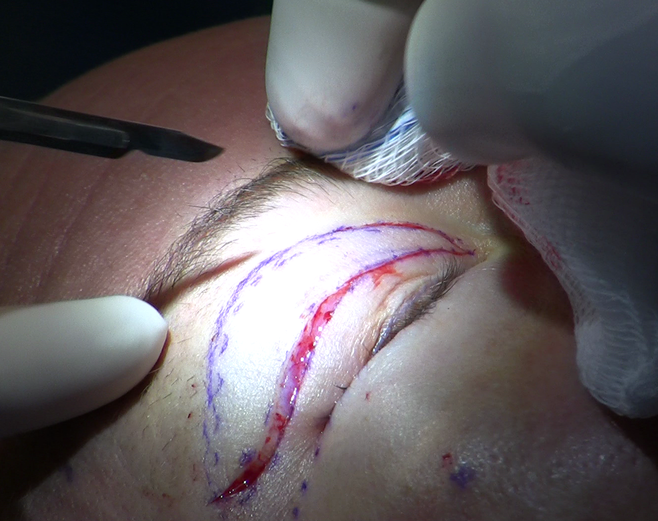
- The surgeon marks the planned crease line at the agreed height and shape with the patient sitting upright.
- Local anesthetic with epinephrine is injected to numb the lid and limit bleeding.
- A full-thickness incision is made along the marked line through skin and orbicularis muscle.
- A strip of orbicularis muscle is removed to allow the crease to form cleanly at that level.
- The orbital septum is opened and the SMFAT layer is assessed; excess tissue is removed as the specific lid requires.
- The preaponeurotic fat pad is examined; only a conservative amount is removed if the lid is unusually full, or it is left intact.
- The lower skin edge is sutured directly to the levator aponeurosis or tarsal plate, forming the adhesion that will become the crease.
- External sutures close the skin; these are removed at five to seven days.
Step seven is the mechanism that makes this result permanent. The skin is physically anchored to the aponeurosis by scar, so the crease forms with every lid movement. Revision requires breaking down established scar tissue - a more demanding operation than the original.
| Feature | Suture technique | Incisional technique |
|---|---|---|
| Incision | Small punctures only | Full-length crease incision |
| Social recovery | Shorter (days) | Longer (1-2 weeks) |
| SMFAT reduction possible | No | Yes |
| Crease permanence | Lower - fading possible over years | High - scar adhesion holds result |
| Practical reversibility | Only within weeks of surgery | Not reliably reversible |
| Best lid type | Thin, low-fat, younger lids | Heavier lids, any fat volume |
Ptosis and the Epicanthal Fold: Two Conditions That Must Be Identified First
Ptosis
Ptosis - reduced lid elevation caused by a weak or stretched levator muscle - affects an estimated 15 to 25 percent of patients who seek double eyelid surgery. It is not rare, and it is the most consequential condition to miss during planning.
When ptosis is present but unrecognized, the surgeon creates a crease and the patient wakes up with the new fold intact - but the lid still droops. The result is a hooded or sausage-like appearance that crease revision cannot fix. The crease is placed correctly. The problem is the levator muscle, and correcting it requires a separate ptosis repair operation targeting that structure specifically.
Measuring ptosis requires a simple clinical test - quantifying lid elevation against a light source in a controlled setting - that should be part of every pre-operative consultation. If your consultation does not include this step, ask for it.
The Epicanthal Fold
The epicanthal fold is the skin fold covering the inner corner of the eye. When this fold is prominent, it physically blocks the new crease from reaching the medial corner. A crease that terminates abruptly mid-lid looks unfinished - particularly with a parallel crease design, which by definition should run the full lid width.
Medial epicanthoplasty releases or reduces the fold so the crease can extend to the inner corner. In one published series of 252 epicanthoplasty cases, 220 - 87 percent - were performed at the same time as double eyelid surgery. That figure reflects how often the conditions coincide and how rarely the decision logic is explained clearly to patients.
Not every patient with an epicanthal fold needs epicanthoplasty. A tapered crease, which naturally narrows toward the inner corner, can work with a moderate fold. Patients planning a parallel crease will more often need the additional procedure, and that should be part of the pre-operative discussion, not a surprise.
Recovery: A Week-by-Week Timeline
- Days 1-3: Swelling peaks. The incisional technique produces more swelling than the suture approach. Cold compresses and sleeping with the head elevated reduce accumulation. The lid looks very swollen and the crease appears high - this is normal at this stage.
- Days 5-7: External sutures are removed after the incisional technique. The crease is visible but the lid looks stiff and unnatural. The result at this point is not representative of the final outcome.
- Weeks 2-4: Most social swelling has resolved. Patients return to daily activity. The crease is present but residual firmness in the lid persists.
- Months 1-3: Swelling within the scar continues to resolve. The crease softens and settles. Most patients reach a stable appearance by three months.
- Months 3-6: Final result. The scar matures, the lid softens fully, and the crease takes on its long-term character. Asymmetry that persists past six months is unlikely to resolve without intervention.
Double eyelid surgery is one of the most frequently performed cosmetic eyelid procedures worldwide, driven by the large proportion of East and Southeast Asian individuals born with a single eyelid.American Academy of Ophthalmology, EyeWiki
Complications Specific to Double Eyelid Surgery
Sausage Eyelid
The sausage eyelid describes a specific outcome in which the pretarsal skin below the crease bulges outward, giving the lid a cylindrical, rolled look. Three separate causes can produce it:
- The crease was set too high, leaving a long pretarsal skin segment that cannot retract properly
- The SMFAT layer was not reduced adequately, so residual bulk pushes the skin forward from beneath
- Unrecognized ptosis was not corrected, and the drooping lid forces the pretarsal skin to bunch below the crease
Identifying which cause - or combination of causes - is driving the appearance determines what revision surgery should address. Targeting only one cause when two are present will produce an incomplete result.
Crease Loss
Crease fading is more common after the suture technique, particularly in heavier lids. Repeated levator contraction works against buried suture anchors over months to years. After the incisional technique, crease loss is less common but can occur if the skin-aponeurosis adhesion is disrupted by significant swelling, trauma, or infection in the early healing period.
Asymmetry
Crease asymmetry is the most common patient complaint after double eyelid surgery. The most frequent underlying cause is a pre-existing difference in levator muscle strength between the two sides that was not measured and addressed during planning. A crease cannot compensate for unequal lid elevation. When one levator is stronger, that lid lifts higher and exposes more of the crease, producing an apparent height difference even when crease placement was technically identical on both sides.
Revision Surgery: What a Second Operation Can and Cannot Fix
Revision after double eyelid surgery is technically more demanding than the first operation. Scar tissue has replaced the normal tissue planes the surgeon worked with originally, and the anatomy is less predictable.
Revision can reasonably address:
- A crease set too high - it can be lowered by releasing the adhesion and re-anchoring at a lower level, though this is technically demanding
- Crease loss after the suture technique - an incisional approach can recreate a stable crease
- Sausage eyelid from inadequate SMFAT reduction - the layer can be addressed through a revision incision
- Asymmetry from crease placement error - the lower crease can be revised upward
- Ptosis missed at the original operation - levator surgery can be added
Revision struggles more with asymmetry caused by pre-existing levator strength differences that were not corrected initially, because the tissue has already been altered and establishing a new baseline is harder. Overcorrection - a crease set so high that extensive scar has formed across a wide area - is also difficult to reverse reliably.
Timing matters. Most surgeons recommend waiting at least six months after the original operation before assessing for revision. Scar tissue continues to mature and soften through that period, and what appears to be a permanent problem at two months may partially resolve by month six.
Frequently Asked Questions
Can the suture technique give me a permanent crease?
It can produce a long-lasting crease, but permanence is not guaranteed. Buried suture anchors are subject to gradual loosening from repeated levator contraction - particularly in thicker or heavier lids. The incisional technique creates a crease maintained by scar adhesion and is considered permanent. The trade-off is that it is also not practically reversible.
What does a 'natural-looking' crease height actually mean in numbers?
For most surgeons, a natural Asian crease falls in the range of 3 to 5.5 mm from the lash line for male patients and 5 to 7.5 mm for female patients. Creases set above that range tend to read as disproportionate on Asian lid anatomy. Ask your surgeon to mark the planned height on your lid with you sitting upright so you can evaluate it before committing.
Do I need epicanthoplasty just because I have an epicanthal fold?
Not automatically. A tapered crease, which narrows toward the inner corner, can work well with a moderate fold. If you want a parallel crease that runs at consistent height across the full lid width, a prominent fold may prevent the crease from reaching the medial corner, and epicanthoplasty becomes worth discussing. Your surgeon should assess the fold as a standard part of pre-operative planning.
How do I know whether I have ptosis that needs treatment at the same operation?
Ptosis presents as a lid that sits lower than it should relative to the pupil, with limited upward excursion. A surgeon can measure this with a brief clinical test at your consultation. If it is not identified and corrected at the same operation, the crease may be placed perfectly but the lid will still droop - and that cannot be fixed by crease revision.
Why do surgeons avoid removing all the fat during this procedure?
Because aggressive preaponeurotic fat removal hollows the upper eyelid in a way that looks skeletal rather than refreshed. This is one of the most cited reasons patients seek revision. When fat reduction is needed in Asian lids, it is the SMFAT layer - the submuscular fibroadipose tissue responsible for the lid's characteristic fullness - that is typically targeted, not the deeper preaponeurotic pad.
What if I regret the suture technique result - can the surgeon just remove the sutures?
Only in the very early post-operative period, before scar tissue has formed. Once scar tissue develops - typically within weeks to a few months - suture removal does not reliably erase the crease. Patients who want to reverse a suture-technique result beyond that point will need to discuss revision surgery, which involves the same scar-based challenges as any crease revision.
General information only, not medical advice. Individual anatomy and healing vary. See the disclaimer.