At some point in the first or second week after lower blepharoplasty, some patients look in the mirror and find something alarming: the white of the eye is no longer flat. Instead, a translucent, blister-like mass pushes forward from under the lower lid, filled with pale, jelly-like fluid. Surgeons call it chemosis. The reassurance most patients receive - "it usually goes away, use lubricating drops" - is true as far as it goes. But it leaves out the mechanism, the grading system, the treatment options, and the logic connecting all three. This guide fills those gaps.
What Chemosis Actually Is
The surface of the eye is lined with a thin, transparent membrane called the conjunctiva. The portion that covers the white of the eye - the sclera - is the bulbar conjunctiva. Directly beneath it lies a second layer called Tenon's capsule, a fibrous sheath that anchors the eye muscles and surrounds the globe. In a healthy eye these two layers lie flush against each other. No visible gap exists between them.
Threaded through the conjunctiva and surrounding tissue is a fine network of lymphatic channels. Their job is to collect fluid that leaks from local blood vessels and carry it away before it accumulates. When that system functions normally, the conjunctiva stays flat and essentially invisible.
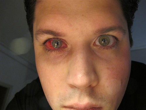
Chemosis occurs when fluid accumulates in the space between the conjunctiva and Tenon's capsule faster than the lymphatic network removes it. The medical term is transudative edema - fluid seeping through blood vessel walls, not pus from an infection or blood from a rupture. The result is that water-blister appearance: a dome of clear membrane filled with straw-colored fluid, visibly raised above the eye's surface. Because the bulbar conjunctiva is transparent, you can often see the fluid beneath it directly. The membrane is not torn, not infected, and not permanently altered. It is distended by trapped fluid with nowhere to drain.

Why Blepharoplasty Causes It
Lower blepharoplasty - whether approached through the skin (transcutaneous) or from inside the lid (transconjunctival) - requires working near the lower conjunctival fornix, the fold where the inner lid lining meets the bulbar conjunctiva. This is where the lymphatic channels of the lower eyelid converge before draining outward.
Dissecting through fat pads, applying electrocautery to seal bleeding vessels, and anchoring the outer corner of the eye during canthal procedures all produce collateral effects on those nearby channels. Channels are cut, sealed with heat, or stretched by retraction. After surgery they cannot drain effectively. Fluid that would normally be cleared in minutes instead pools in the subconjunctival space.
This is not a technical error. It is a predictable consequence of operating in tissue that depends on lymphatic continuity. The network will gradually re-establish drainage, but the timeline is weeks in most patients and can stretch to months in others.
Upper blepharoplasty causes chemosis far less often. The upper lid's conjunctival anatomy sits farther from the dissection planes used for lower-lid fat removal and canthal work. That anatomical gap explains why chemosis is predominantly a lower-lid problem.
Risk Factors That Raise the Odds
Published series have found chemosis after lower blepharoplasty to be considerably more common than the figure typically quoted in pre-operative consultations - some surgeon series report it in more than one in four patients. Several factors push individual risk higher.
Patient-level factors
- Preexisting dry eye syndrome - the conjunctiva is already mildly inflamed before surgery, and the compromised baseline makes post-operative fluid accumulation harder to clear
- Thyroid eye disease - chronic orbital and conjunctival inflammation that amplifies the surgical response
- Significant eyelid laxity or scleral show - lower-lid anatomy in these patients reduces structural containment of accumulating fluid
- Hormone therapy use - alters fluid-handling and healing dynamics at the tissue level
- Combined upper and lower blepharoplasty in one session - increases total tissue disruption, operative time, and the cumulative burden on periorbital lymphatic drainage
Surgical factors
- Transcutaneous lower blepharoplasty combined with lateral canthoplasty - head-to-head published comparisons show a statistically significant higher rate of persistent chemosis versus a transconjunctival approach with canthopexy; this is the highest-risk surgical combination
- Longer operative time - more time in the field means more cumulative tissue handling and greater lymphatic disruption
- Any lateral canthal procedure - the outer canthal angle is lymphatic-rich; both canthoplasty and canthopexy bring instruments into direct proximity with those channels
Patients undergoing the transcutaneous approach plus lateral canthoplasty should receive explicit pre-operative counseling that chemosis is a common, manageable outcome - not a rare complication that signals something wrong.
Grading Severity: What Each Level Looks and Feels Like
A published four-point classification system describes chemosis by how it affects lid function and how long it persists. Knowing where a case falls tells both patient and surgeon which treatment tier is appropriate.
| Grade | Appearance and lid function | Timeline marker | Clinical priority |
|---|---|---|---|
| Grade 1 | Mild elevation; lids close fully | Typically resolves within 2-3 weeks | Conservative management |
| Grade 2 | Pronounced swelling; lids cannot fully close | Acute, often first 1-2 weeks post-op | Prompt escalation to topical steroids |
| Grade 3 | Persists beyond 3 weeks despite treatment | Subchronic, ongoing | Active reassessment; consider conjunctivoplasty |
| Grade 4 | Chemosis with lower lid malposition | Variable | Surgical evaluation strongly indicated |
Chemosis is frequently confused with other post-blepharoplasty findings. The distinction matters because treatments differ entirely.
- Chemosis - the conjunctival membrane itself is swollen; the eye surface looks wet and blister-like; the lid margin may be in normal position; not typically very painful; no discharge
- Lower eyelid retraction - the lid margin has pulled downward, exposing sclera below the iris; the conjunctiva is flat; this is a structural lid problem, not a conjunctival one
- Ectropion - the lower lid margin rolls outward; the inner lid surface becomes visible; a mechanical lid problem requiring different management
- Conjunctivitis or infection - painful, red, with mucopurulent discharge; chemosis produces no pus and causes discomfort rather than acute pain
Grade 4 is the one exception where chemosis genuinely mimics lid malposition, because a large herniated mass of conjunctiva can mechanically hold the lower lid away from the globe. Even here the correct first step is treating the chemosis directly - not adding a lid-tightening procedure on top of it.
The Treatment Ladder
Level 1 - Conservative care
Start these steps immediately after surgery and maintain them through at least the first two weeks. This is not passive waiting - it is active management that determines whether mild chemosis resolves quickly or progresses into a harder problem.
- Apply preservative-free lubricating eye drops four to six times daily from the first post-operative day - frequency matters; waiting until the eye feels dry is too infrequent to maintain the protective film
- Apply cold compresses for fifteen to twenty minutes, several times daily, during the first two to three days; cold reduces vascular permeability and slows the rate of fluid accumulation in the subconjunctival space
- Elevate the head during sleep using a wedge pillow or extra pillows; lying flat allows fluid to pool under hydrostatic pressure
- Apply preservative-free ophthalmic ointment at night - the thicker consistency maintains contact with the eye surface through sleep and protects tissue that the closed lid may not fully cover
- If the conjunctiva is herniated over the lid margin, add nighttime lid taping with paper surgical tape, placing a horizontal strip gently across the lower lid to keep lid-to-globe contact through the night
Level 2 - Topical corticosteroids
When conservative measures have been correctly applied for a week or more without adequate improvement, a short course of topical corticosteroid eye drops can interrupt the inflammatory cycle. Agents in the prednisolone and loteprednol families are commonly used for a five-to-seven-day course. These require a prescription. Do not start steroid drops without direct guidance from your surgeon or an ophthalmologist - they carry real risks in certain eye conditions, and the diagnosis needs to be confirmed first.
Level 3 - Snip conjunctivoplasty
When chemosis reaches Grade 3 or 4 - persisting beyond three weeks despite active treatment - an in-office procedure can resolve it quickly. The snip conjunctivoplasty uses small scissors to excise a narrow strip of swollen conjunctiva and underlying Tenon's capsule. The trapped fluid releases immediately. The blister deflates. Surrounding tissue heals within days.
This does not require a return to a hospital operating room. It is performed in an office or outpatient setting under topical anesthetic drops, with local injection available if needed. Published case series report complete resolution at one-week follow-up with no recurrence at six months. The procedure was formally described in the surgical literature by Jones, Georgescu, McCann, and Anderson in a 2010 paper in Archives of Facial Plastic Surgery - it has a well-documented history spanning more than fifteen years. For patients who have spent weeks managing severe chemosis and assumed nothing remained except waiting, this option is the most practically useful information available to them.
Week-by-Week Recovery: Normal Progress and Red Flags
Week one: Some chemosis is normal and expected after lower blepharoplasty. The conjunctiva may be visibly elevated, the lids may feel stiff, and the eye may water more than usual. Cold compresses and lubricants should be in use from day one. Mild swelling in this window, managed actively, almost always resolves without escalation.
Week two: Grade 1 cases should show measurable improvement - smaller blister, easier lid closure, improving comfort. Cases not improving by day ten to fourteen warrant contact with the surgical team. This is the window for initiating topical steroids if the conservative level has not worked.
Week three: Any chemosis still present at three weeks crosses into Grade 3. Passive observation beyond this point is not appropriate management. The surgical team should formally reassess and determine whether snip conjunctivoplasty is indicated.
Weeks four to six: Most patients who received prompt, aggressive early management reach baseline comfort and visual function within this window. Patients with preexisting dry eye or thyroid eye disease, or those who delayed treatment, may take longer.
Contact the surgical team promptly - not at the next scheduled appointment - if any of these occur:
- Pain escalating rather than staying steady or improving
- Yellow or green discharge from the eye
- Any reduction in vision
- Chemosis worsening after the first week rather than stabilizing
- The lower lid visibly drooping or rolling outward
What Patients Can Do at Home Right Now
Use preservative-free artificial tears during the day - the preservatives in standard multi-dose bottles can irritate already-stressed conjunctival tissue and worsen the condition. At night, switch to a preservative-free ophthalmic gel or ointment; the thicker viscosity stays in contact longer. Avoid redness-relieving drops that "get the red out" - the vasoconstrictors in them cause rebound dilation and compound the underlying problem.
For lid taping, use paper surgical tape from any pharmacy - never regular adhesive tape on eyelid skin. Apply a short horizontal strip across the lower lid to maintain lid-globe contact. Apply ointment to the eye first, then close gently before taping. On waking, re-lubricate before removing the tape to prevent pulling.
Several things actively delay recovery and are worth eliminating for the first two weeks:
- Rubbing the eye - mechanical pressure on herniated conjunctiva can worsen the blister and disrupt healing
- High-sodium foods and alcohol - both promote systemic fluid retention that redistributes to swollen tissue
- Strenuous exercise - elevated blood pressure increases transudation from periorbital vessels
- Contact lens use - until the surgeon confirms the conjunctival surface is fully healed
- Sleeping flat - even one night without head elevation can undo several days of progress in early-stage chemosis
How Chemosis Connects to Other Post-Blepharoplasty Problems
Dry eye can worsen meaningfully. The goblet cells embedded in the conjunctiva produce the mucin layer of the tear film - the layer that stabilizes the tear film and allows tears to spread and adhere evenly to the corneal surface. Persistent chemosis stresses those cells directly. Patients with marginal pre-operative dry eye can leave the chemosis episode with a lasting reduction in tear quality that outlasts the swelling itself by months.
Corneal exposure becomes a genuine injury risk at Grade 2 and above. When the lower lid cannot fully close, the central cornea loses the blink reflex and continuous tear film contact. The corneal epithelium can break down within days under those conditions. This is why lubricant frequency and nighttime ointment coverage are not optional steps - they are protecting the cornea, not just the conjunctiva.
Lid malposition can follow from Grade 4 chemosis left untreated long enough. A large conjunctival mass holding the lid away from the globe, sustained over weeks, can distort the anatomical relationships that keep the lid in position. Treating the chemosis early prevents this trajectory and avoids the need for a second corrective procedure later.
Chemosis after blepharoplasty is predictable, manageable, and fully reversible when treated through the right steps at the right time. What converts a common post-operative finding into a prolonged problem is delay - delay in starting lubricants, delay in escalating to steroids, and delay in recognizing when the office procedure is the answer.
Frequently Asked Questions
Is chemosis a sign that my surgeon made a mistake?
Not in most cases. Chemosis is a predictable result of operating near conjunctival lymphatic channels - disrupting those channels is inherent to lower eyelid dissection, not a technical error. Surgical choices do affect risk, though: the combination of a transcutaneous approach with lateral canthoplasty carries statistically higher risk than less-invasive alternatives, and patients deserve to know that before selecting an approach.
How long does chemosis typically last after eyelid surgery?
Mild cases treated promptly from day one typically resolve within two to four weeks. Moderate to severe cases, or those in patients with preexisting dry eye or thyroid disease, can persist for several months if treatment is delayed or insufficient. The single largest variable in duration is how quickly active management begins.
Can chemosis permanently damage my eye?
When managed appropriately, chemosis resolves without permanent consequences. The risk of lasting damage arises when it goes untreated long enough to produce corneal exposure injury - the surface of the eye can break down when the lid cannot fully close over it. Grade 2 and above requires prompt attention rather than passive observation for this reason.
What exactly is the difference between chemosis and ectropion?
Chemosis involves the conjunctival membrane on the eyeball surface - it looks like a wet blister on the white of the eye. Ectropion is a structural problem with the lower eyelid itself, where the lid margin rolls outward and the inner lid surface becomes visible. They are anatomically distinct and require different treatments. Grade 4 chemosis can appear to cause lid malposition because the swollen mass holds the lid away from the globe, but treating the chemosis directly is the correct first step before considering any lid procedure.
When should I call my surgeon instead of just waiting?
Call rather than wait if the swelling is increasing after the first week, if the lids cannot fully close, if you notice any change in vision, or if pain is escalating. Also call at the three-week mark regardless of how the eye looks - that threshold marks the transition to subchronic chemosis, which requires formal reassessment and possible escalation to the in-office snip procedure.
Is the snip conjunctivoplasty procedure painful?
Topical anesthetic drops numb the eye surface before the procedure begins, and local anesthesia can be added for additional comfort. Most patients report minimal discomfort during the procedure itself. Published case results describe complete resolution by the one-week follow-up, with no recurrence at six months - and the procedure is performed in an office setting, not an operating room.
General information only, not medical advice. Individual anatomy and healing vary. See the disclaimer.