If you have been told you have under-eye bags and treated accordingly - filler injections, creams, maybe a blepharoplasty consultation that went nowhere - there is a specific reason the problem persisted. You may have festoons: a structurally different condition that sits below the eyelid itself, involves different tissue layers, and fails to respond to treatments designed for the other two under-eye conditions. Getting the diagnosis wrong is common. Getting the treatment wrong, especially with filler, can make the condition measurably worse.
Two other articles on this site cover herniated fat-pad bags and tear-trough hollows. Festoons are the third entity in that trio - the one most frequently misdiagnosed and most often worsened by well-intentioned intervention.
What Festoons Actually Are: Anatomy in Plain Language
The orbicularis oculi is the ring-shaped muscle that encircles the eye and controls blinking. Just above the cheekbone, a ligament called the orbicularis retaining ligament - also called the orbitomalar ligament - anchors this muscle and the overlying skin to the underlying bone. First formally described by Kikkawa and colleagues in 1996, this ligament forms the upper boundary of a small anatomical compartment called the prezygomatic space: a potential pocket sitting between the muscle and the cheekbone, just below and outside the lower eyelid.
The floor of this compartment is the zygomatico-cutaneous ligament, which is considerably stronger and maintains its integrity with age. The ceiling - the orbicularis retaining ligament - weakens progressively. The result is a compartment whose top boundary becomes increasingly slack while the bottom holds firm. Lymphatic fluid, which should circulate freely through this area, begins to pool. It collects in a predictable crescent shape across the upper cheek, draped between the inner and outer corners of the eye. That crescent is a festoon.
This is entirely different from the fat-pad bag, which involves orbital fat pushing forward through a weakening orbital septum inside the eyelid itself. Festoons do not primarily contain orbital fat. They are a combination of lax skin, lax orbicularis muscle, and trapped lymphatic fluid in a structurally compromised space. Treating orbital fat leaves all three of those components untouched.
Festoons vs. Malar Bags: A Meaningful Clinical Distinction
The two terms are frequently used interchangeably, but they describe different points on the same spectrum, and the distinction has direct implications for what treatment will work.
Malar bags are the earlier, milder form. The tissue has not yet developed the visible draping and structural laxity of a full festoon. The swelling is predominantly fluid and tends to fluctuate: most pronounced first thing in the morning, softer by midday as gravity and upright movement help lymph drain. Patients with malar bags often describe looking puffy in early-morning photographs but more normal by afternoon.
Established festoons have advanced to the point where the skin and muscle fold is visible and relatively persistent regardless of time of day or body position. Some fluctuation may still occur, but the structural drape - the actual redundant tissue - cannot be resolved by improving drainage alone. That distinction is what separates a condition where non-surgical treatment can genuinely help from one where surgery is the only reliable path.
| Feature | Fat-Pad Bags | Festoons / Malar Bags | Tear-Trough Hollows |
|---|---|---|---|
| Primary tissue | Herniated orbital fat | Lax skin, muscle, trapped fluid | Volume loss, soft tissue deficit |
| Location | Below lid, inside orbital rim | Upper cheek, below orbital rim | Inner under-eye, along orbital rim |
| Fluctuates with posture? | No | Yes - worse lying flat | No |
| Texture on palpation | Firm, immovable | Soft, compressible, shiftable | Sunken, no resistance |
| HA filler appropriate? | Sometimes, with care | No - actively worsens the condition | Yes, often first-line |
| Surgical gold standard | Transconjunctival blepharoplasty | Direct excision with canthopexy | Fat repositioning or volume restoration |
Three At-Home Self-Checks Before Your Consultation
None of these replace clinical examination. But they sharpen your observations and may spare you from undergoing treatment aimed at the wrong condition.
The Touch Test
Press the puffy area gently with one fingertip. Fat-pad bags feel firm and immovable - what you are pressing against is fat contained within the orbit. Festoon tissue feels soft and somewhat compressible. You may be able to shift it slightly with gentle lateral pressure. That softness and fluctuance reflects lymphatic fluid pooling in loose tissue, not herniated fat.
The Time-of-Day Test
Check the area before getting up in the morning, then again in the mid-afternoon. True fat-pad bags are essentially constant throughout the day - fat does not drain. Festoon-related swelling is typically most pronounced on waking, after a night of horizontal positioning in which lymph has been unable to drain effectively, and softens somewhat once you have been upright and moving for a few hours.
The Position Test
If you spend time lying flat - reading, watching television - and notice the puffiness worsening, that is consistent with festoon behavior. Gravity assists lymphatic return when you are upright; it stops helping when you are horizontal. This postural sensitivity does not apply to fixed fat-pad bags.
What Causes Festoons to Develop
Festoon formation is a convergence of several processes. Identifying which ones are active matters, because some are addressable before any treatment of the festoon itself begins.
- Genetics and ligament laxity: Inherited weakness of the orbicularis retaining ligament and naturally thin skin accelerate the entire process. Some patients develop visible festoons in their 30s, well before other significant facial aging appears, because of this baseline anatomical disadvantage.
- Cumulative UV exposure: Collagen and elastin degradation from sun damage compounds skin and muscle laxity over time. UV exposure is the most modifiable long-term risk factor for early festoon development.
- Age-related muscle weakening: The orbicularis oculi loses tone and structural integrity with age, making it increasingly likely that the tissue will drape rather than hold its contour.
- Smoking and chronic dehydration: Both accelerate collagen breakdown and impair tissue quality, worsening the underlying structural laxity.
- Systemic health conditions: This is almost never mentioned in patient-facing information, and it matters significantly. Hepatic disease, cardiac failure, renal disease, hypothyroidism, and chronic allergies can all cause or substantially worsen malar edema. A thorough medical history to rule these out is a prerequisite before attributing festoons to anatomy alone. If a systemic condition is driving the fluid retention, no amount of local treatment will produce lasting improvement.
If your festoons appeared suddenly, worsened rapidly, or are accompanied by swelling elsewhere in the body, discuss this with a physician before pursuing aesthetic intervention.
The Filler Trap: Why Hyaluronic Acid Makes Festoons Worse
This is the mechanism behind the most preventable harm in festoon patients, and it is worth understanding precisely rather than accepting as vague "contraindication" language.
Hyaluronic acid - the material in most soft-tissue fillers - is hydrophilic. It binds water. In a tear trough, where the goal is volume replacement, that property is useful. In a festoon, it is harmful for two distinct reasons. First, by attracting water, an HA filler draws additional fluid into a space that already has compromised lymphatic drainage, increasing the fluid load on a system that is already failing to clear it. Second, the physical volume of the injected material compresses local lymphatic channels. Lymphatics are thin-walled and low-pressure; adjacent compression impairs their function mechanically, independent of the hydrophilic effect. Together, the two mechanisms create a self-reinforcing cycle: the filler attracts more fluid, which compresses more lymphatics, which allows more fluid to accumulate.
The prezygomatic space has a compromised lymphatic architecture even before any treatment. Anything that adds volume or increases fluid load within it amplifies rather than addresses the underlying problem.Mechanism described in published oculoplastic literature and clinical guidance from festoon specialists
If Filler Has Already Been Injected
Filler-induced festoon worsening may persist even after hyaluronidase dissolution, because the filler has already disrupted local lymphatic architecture. Despite this, early dissolution remains the recommended first step - acting quickly gives the tissue the best chance of recovery. If you are in this situation, take these steps in order:
- Stop any further filler treatments to the under-eye or upper cheek area immediately.
- Contact the practitioner who administered the filler and describe the worsening directly.
- Request hyaluronidase dissolution promptly - earlier treatment is associated with better outcomes than waiting for the filler to resorb on its own.
- Allow the tissue several weeks to settle before drawing conclusions about residual appearance.
- Consult a specialist experienced specifically in festoons before deciding on any next steps.
Non-Surgical Treatment Options and What to Expect From Them
A 2020 systematic review in Aesthetic Surgery Journal found no single non-surgical treatment that meets all the criteria for effective festoon management. That is an honest appraisal of the current evidence. There is, however, a meaningful treatment ladder for mild-to-moderate cases before surgery becomes the only option.
Sclerotherapy: Doxycycline and Tetracycline Injections
Despite being the most evidence-supported non-surgical approach, intralesional sclerotherapy is almost invisible in patient-facing content on festoons. The technique involves injecting doxycycline or a related antibiotic directly into the festoon compartment. The mechanism is controlled inflammatory fibrosis: the injection provokes a healing response that collapses the fluid pocket and reattaches the loose tissue layers. A published preliminary study by Goldberg and colleagues documented clinically significant reductions in festoon grade following this treatment, with no dermatological or visual complications. A 2021 study in Aesthetic Surgery Journal Open Forum combined intralesional tetracycline with a pinch-technique skin excision and a lateral canthopexy procedure, reporting marked improvement in severe festoons with limited visible scarring compared to excision alone. Results are not immediate; the fibrotic remodeling takes weeks to mature.
CO2 Laser and Radiofrequency
CO2 laser resurfacing removes damaged surface skin and stimulates collagen remodeling, tightening the overlying tissue. It is rarely sufficient to resolve festoons on its own, but is commonly used alongside surgical excision to improve overall skin quality and reduce scar visibility. Radiofrequency devices that heat the deeper dermis work by collagen contraction and are sometimes used in early-stage cases where skin laxity is mild. Neither approach addresses structural muscle or ligament laxity directly; expectations for standalone use should be conservative.
Surgical Treatment: Excision, Canthopexy, and Combined Approaches
For severe or persistent festoons with significant structural laxity, surgery is the most reliable path to lasting correction. Understanding what the procedure involves - and what its limits are - helps set realistic expectations before consultation.
Assessing Lower Eyelid Laxity First
Before any festoon surgery, the surgeon will assess lower eyelid support. The snap-back test works by gently pulling the lower lid away from the eye and observing how quickly and fully it returns to its resting position. A well-supported lid returns immediately. With increasing laxity, return slows; in more advanced cases the lid remains displaced until the patient deliberately blinks. Patients at the midpoint of the laxity scale or beyond typically need a concurrent lid-tightening procedure - lateral canthopexy or canthoplasty - alongside festoon surgery. Without it, the lower lid can be pulled downward by postoperative swelling and scarring. This is far easier to prevent than to correct after the fact.
Direct Excision
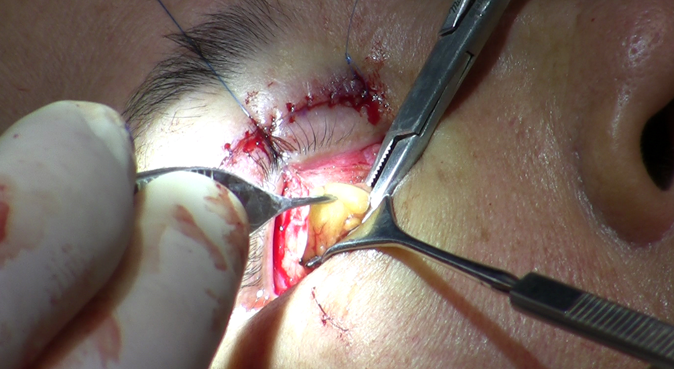
The definitive technique involves marking the festoon tissue precisely, excising it, and closing the wound with sutures placed below the eyelid and across the upper cheek. This provides the greatest degree of correction for severe cases. The trade-off is scar visibility, which varies substantially depending on patient skin type, individual healing, and surgical precision. For patients with poor scar history or particularly pale and thin skin, this trade-off deserves a candid pre-operative discussion. For many, the scar - which typically sits in a natural skin fold - is an acceptable exchange for resolution of a problem that has not responded to anything else.
Combined Approaches for Better Outcomes
Contemporary festoon surgery rarely relies on excision alone. Combining techniques produces better outcomes with lower recurrence. A typical combined approach may include:
- Direct excision of the festoon tissue with careful wound closure
- Lateral canthopexy or canthoplasty to support the lower eyelid and prevent post-operative malposition
- Intralesional sclerotherapy to further collapse the compartment and reduce the likelihood of fluid reaccumulation
- Adjunctive CO2 laser resurfacing to smooth skin texture and soften the appearance of the closed incision
Recovery from festoon excision typically involves significant bruising and swelling for the first week to ten days, followed by a longer period of residual firmness as the scar matures. Final results become visible at several months, though individual healing timelines vary.
Choosing the Right Specialist
Festoon surgery sits in a narrow anatomical territory: it touches the lower eyelid, the orbital rim, and the upper cheek simultaneously. Oculoplastic surgeons - who train in both ophthalmology and plastic reconstructive surgery of the eyelid and orbit - have specific familiarity with the eyelid mechanics that must be preserved during the procedure. General plastic surgeons with documented and repeated experience treating festoons are also appropriate candidates. The key variable is not the specialty designation but the number of times the surgeon has performed this specific operation - not related procedures, but festoon excision itself.
Useful questions to ask at a consultation:
- How many festoon procedures have you performed, and can I see before-and-after images from your own patients?
- Will you assess my lower eyelid laxity, and if it is significant, what do you plan to do about it alongside the festoon correction?
- Are you recommending sclerotherapy, excision, or a combination - and what is the reasoning for my specific anatomy?
- What does the scar typically look like at six months in patients with skin similar to mine?
- Have you considered whether any systemic health factors may be contributing to my swelling, and should I have a medical workup before proceeding?
A surgeon who answers these questions with specificity rather than generalizing to blepharoplasty or facial rejuvenation broadly is demonstrating genuine familiarity with what festoon correction requires.
Frequently Asked Questions
Can festoons resolve on their own without treatment?
Mild malar bags that are primarily fluid-driven may improve if an underlying contributor - such as allergies, thyroid dysfunction, or sleep positioning - is identified and addressed. Established festoons with structural skin and muscle laxity do not resolve on their own. The changes to the orbicularis retaining ligament and surrounding tissue are not reversible without intervention, though managing systemic factors can prevent further progression.
How is a festoon different from a standard under-eye bag?
Fat-pad bags sit inside the orbital rim and involve orbital fat pushing through a weakening septum. Festoons sit below the orbital rim on the upper cheek and consist of lax skin, lax muscle, and trapped lymphatic fluid - not primarily fat. They are in a different location, involve different tissue, and require different treatment. Transconjunctival blepharoplasty, which removes or repositions orbital fat, does not address festoon tissue at all.
How can I tell if my filler made my festoons worse?
If filler was placed near the under-eye or upper cheek area and the puffiness increased rather than improved - and has not resolved with time - this pattern is consistent with filler-aggravated festoons. The swelling is typically soft, somewhat variable with posture, and may cover a broader area than what was originally treated. Contact the practitioner who administered the filler promptly; earlier hyaluronidase dissolution is associated with better outcomes than waiting for the material to resorb naturally.
Is there any non-surgical option that actually works?
For mild festoons and malar bags where fluid accumulation is the dominant component, intralesional sclerotherapy with doxycycline or tetracycline has published evidence of meaningful improvement and is significantly underused in clinical practice. Skin-tightening procedures can add incremental benefit for surface laxity. Established festoons with visible tissue draping have not been shown to fully resolve with non-surgical approaches alone - a surgical consultation is warranted to understand the full picture before committing to either path.
What is the snap-back test, and why does my surgeon want to do it?
The snap-back test measures how well your lower eyelid returns to its normal position after being gently pulled away from the eye. It tells the surgeon whether your lid has enough structural support to tolerate festoon surgery safely, or whether a lid-tightening procedure needs to be added to prevent the lower lid from drooping or pulling outward as the surgical site heals. Skipping this assessment and proceeding without addressing significant laxity can result in a complication that requires a second operation to correct.
Do festoons come back after surgery?
Recurrence is possible, particularly when the underlying tendency toward ligament laxity is strong. Combined approaches - combining excision with sclerotherapy and canthopexy - appear to reduce recurrence compared to excision alone based on published outcomes. Long-term habits matter too: consistent sun protection, not smoking, and controlling any systemic health factors that contribute to fluid retention give surgical results the best chance of holding over time.
General information only, not medical advice. Individual anatomy and healing vary. See the disclaimer.