Patients who have upper blepharoplasty to look rested can sometimes come out looking gaunt instead. The crease is crisp, the excess skin is gone, but the space beneath the brow has caved inward and a shadow now runs along the upper orbit that was never there before. This is surgical hollowing - a specific consequence of volume loss that has a precise anatomical explanation, a set of identifiable risk factors, and multiple treatment pathways with genuinely different profiles. Understanding which path fits depends on knowing exactly what was lost and where it sat.
What a Hollowed Upper Eyelid Looks Like
The central visual problem is a shadow. When fat volume is removed from the upper eyelid, the bony orbital rim becomes prominent relative to the globe, and a triangular dark area forms in the sulcus - the groove between the brow and the upper lid margin. Surgeons call this the A-frame deformity. The orbital rim functions like the ridge of a tent frame; without soft tissue filling the space beneath it, light falls across the bone and casts a deepening shadow downward into the eyelid. The eyelid does not look refreshed. It looks hollow.
Overhead lighting makes it dramatically worse. Ceiling fixtures, outdoor midday sun, fluorescent office lights - any source that projects from directly above sends light across the bony rim and intensifies the shadow below it. A useful self-assessment is to stand under a ceiling light and look in a mirror. If a pronounced shadow sits in the upper sulcus, particularly one that was not there before surgery, that is the A-frame shadow. Photographs taken under consistent overhead light at different points over time document whether the hollowing is stable or progressing.
- The sulcus appears deeper than before surgery, not shallower
- The bony rim of the orbit looks prominent or skeletal rather than soft
- The shadow worsens under overhead light and improves under diffuse frontal light
- The eye reads as sunken or exhausted rather than alert
- The appearance has continued to worsen gradually since surgery
Patients often describe the result as looking 'overdone' precisely because fullness in the upper eyelid is associated with youth and health. A hollow, skeletonized orbit communicates illness or advanced age in a way that the original sagging skin never did.
Anatomy Behind the Crease: Two Fat Pads and the ROOF
The two orbital fat compartments
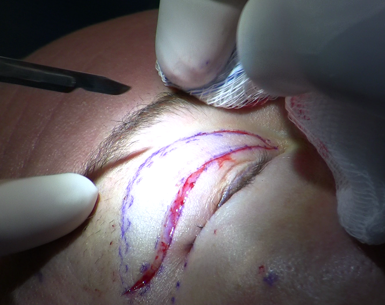
The upper eyelid contains two distinct fat compartments with different locations, appearances, and surgical significance. The central fat pad - also called the preaponeurotic fat pad - sits just anterior to the levator aponeurosis, the tendon responsible for lifting the eyelid. This is the largest, most surgically accessible compartment and the one most commonly targeted during blepharoplasty. The medial fat pad, also called the nasal fat pad, sits closer to the nose and is separated from the central pad by the interpad septum, a thin membrane formed by an extension of Whitnall's ligament. The nasal pad is visually distinct in surgery: it is noticeably whiter in colour than the central pad, which helps experienced surgeons identify it separately rather than treating the two compartments as one continuous mass to be excised.
The ROOF layer
A third layer sits separately from both orbital fat pads: the retro-orbicularis oculi fat, or ROOF. This layer lies deep to the orbicularis muscle but superficial to the orbital rim, contributing independently to the contour of the brow and the upper sulcus. The ROOF is not part of the orbital fat compartments and is not the same tissue that surgeons remove during standard blepharoplasty. Its atrophy is one of the primary drivers of upper eyelid hollowing in both surgical and non-surgical patients - a fact that treatment approaches ignoring this layer will miss entirely.
- Central preaponeurotic fat pad: provides the main visible bulk of the upper sulcus; most commonly over-removed during surgery
- Medial nasal fat pad: contributes to nasal sulcus volume; whiter in colour; can be repositioned centrally rather than excised
- ROOF layer: separate from both pads; sits beneath the orbicularis; a distinct volume contributor that depletes with age and is rarely addressed in standard blepharoplasty
How Over-Removal Happens
For most of blepharoplasty's surgical history, fat removal was the explicit aim. A full upper sulcus was treated as excess tissue, and aggressive excision of the preaponeurotic fat pad was considered thorough technique. This approach was reinforced by how clearly the fat presented during surgery: the pad is prominent, easy to access once the orbital septum is opened, and removing it creates an immediately visible change in eyelid contour on the table.
The problem is that the tissue providing bulk in youth also provides structural support. Remove too much of it and the orbital rim becomes skeletal. The change is not always immediate: in the months after surgery, residual swelling can mask mild hollowing. The true settled contour emerges only after the eyelid heals fully - often six to twelve months post-operatively.
Surgical thinking has shifted significantly toward volume conservation and redistribution. A 2011 published series by Massry in Ophthalmic Plastic and Reconstructive Surgery documented a nasal fat pad repositioning technique - sliding the medial fat pad centrally rather than excising it - and found zero cases of new or worsening superior sulcus hollowing at follow-up. That result has shaped how careful practitioners approach the procedure now. Patients who were operated on during the earlier era of aggressive excision are precisely those who present years later seeking revision.
Volume redistribution rather than removal has become the direction upper eyelid surgery has moved toward - because the consequences of over-removal grow more visible and harder to reverse as the patient ages, while repositioned fat maintains its volume without the resorption uncertainty of grafted tissue.
Who Is at Highest Risk Before Surgery
Not every patient who has upper blepharoplasty develops hollowing, but certain anatomies make it nearly certain unless the surgeon adjusts their approach accordingly. A thorough pre-operative evaluation should identify these before a single incision is made.
- Deep-set or sunken orbits: Patients whose bony orbital margin is already prominent relative to the globe are at significantly higher risk. Even conservative fat removal in these eyes creates visible hollowing because there is so little volume to spare.
- Thin eyelid skin: Fine, translucent upper eyelid skin makes any contour change - whether a depleted sulcus, a lumpy filler result, or a visible fat graft - more apparent to observers.
- Pre-existing age-related volume loss: A patient with meaningful ROOF atrophy and orbital fat depletion already present before surgery has less margin for further removal. Excising additional fat at this stage can push a borderline contour into obvious hollowing.
- High or prominent brow position: A brow sitting high relative to the orbital rim exaggerates the visual depth of the sulcus, making even moderate hollowing appear severe under lighting.
Surgeons who skip orbital depth assessment and treat every upper eyelid as if it has tissue to spare are taking a risk that the patient will bear the consequences of.
Distinguishing Surgical Hollowing from Natural Aging
When a patient had upper blepharoplasty several years ago and now presents with hollow upper eyelids, parsing the cause has direct implications for treatment decisions. Blaming surgery when natural aging is the dominant driver, or the reverse, leads to treatment plans that miss the actual problem.
Natural upper eyelid hollowing begins with ROOF atrophy that starts in the mid-twenties - earlier than most patients realise. The depletion continues gradually each decade, accompanied by slow loss of orbital fat volume. By the time a patient is in their fifties or sixties, meaningful natural hollowing can exist independently of any surgical history. If a blepharoplasty occurred ten years earlier, separating what surgery caused from what time caused becomes genuinely difficult.
- Pre-operative photographs are the most reliable reference: if significant hollowing was already present before surgery, the current appearance reflects both surgical and natural causes
- Surgical hollowing typically becomes apparent as post-operative swelling resolves within the first year; natural hollowing progresses slowly over years or decades
- Hollowing that has continued worsening for five or more years post-operatively almost certainly reflects ongoing ROOF and orbital fat atrophy alongside any original surgical volume loss
- Hollowing concentrated specifically in the central sulcus - where the preaponeurotic fat pad sat - is more consistent with surgical over-removal than generalised natural aging
Post-blepharoplasty hollowing tends to worsen progressively over time because the aging process continues depleting whatever orbital fat and ROOF tissue remains after surgery. Treating it once and expecting the correction to hold indefinitely is not realistic - managing this condition is a long-term process, not a single repair.

Non-Surgical Fix: Hyaluronic Acid Filler
For mild to moderate hollowing, hyaluronic acid filler placed in the correct anatomic planes is the most accessible first-line treatment. Product selection is not interchangeable.
Why the filler type is not negotiable
Firm, high-G-prime hyaluronic acid fillers - appropriate for cheeks or lips where tissue is thick - are wrong for the upper eyelid. Thin eyelid skin stretched over a stiff gel creates visible lumps that are palpable and sometimes visible to others. More visibly, high-G-prime products placed too superficially cause the Tyndall effect: a blue-grey discolouration produced by light scattering through a gel sitting near the skin surface. Low-G-prime products - Belotero Balance, Restylane Silk, Juvéderm Volbella - are preferred because their softer consistency integrates with fine tissue and remains invisible under the skin, even if placement is not perfectly deep.
The dual-plane injection technique
Single-layer injection is insufficient. The dual-plane technique targets two anatomic planes simultaneously: the ROOF layer and the supraorbital fat space. The injector places filler deep to the orbicularis muscle using a threading approach - depositing material in fine linear passes as the needle or cannula withdraws. This deep placement bypasses the superficial layer where Tyndall effect and lumping occur.
A 2024 PMC study of this technique for sunken upper eyelids found corrections maintained at twelve months of follow-up, with no cases of vascular occlusion and no Tyndall effect when injections were consistently placed in the correct deep planes. Technique proved as important as product - the same filler placed one plane too shallow produced exactly the complications the technique was designed to prevent. The reversibility of hyaluronic acid with hyaluronidase is a meaningful safety advantage: an unsatisfactory result can be dissolved and reconsidered.
Surgical Fix: Fat Grafting
When hollowing is moderate to severe, or when a patient wants a longer-lasting structural correction, autologous fat grafting is the standard surgical option. Fat is harvested from the abdomen or inner thigh, processed by centrifugation or decanting to concentrate viable cells, then injected in fine retrograde threads beneath the orbicularis muscle into the depleted sulcus. Only the patient's own tissue is used.
Understanding resorption
Fat grafting in the periorbital area resorbs substantially - often a third to a half of the transferred volume - in the first three to six months as the body absorbs injured cells and the graft vascularises. Surgeons account for this by intentionally overcorrecting at placement, accepting temporary puffiness during the settling period so the final volume lands at the target. A patient who is not warned about this will be alarmed when their eyelids look over-filled for weeks after the procedure.
A 2024 PMC study of severe upper eyelid depression treated with combined orbital fat repositioning and autologous fat transplantation in 79 patients reported high patient satisfaction at one year, a low recurrence rate, and a low overall complication rate. Results like these reflect careful patient selection and experienced technique - they are not universal guarantees. The fat that survives the initial resorption phase tends to remain stable long-term, though ongoing natural aging will continue to reduce total upper eyelid volume over the years.
The Intermediate Option: Orbital Fat Transposition
Between filler and full fat grafting sits a surgical option that is almost entirely absent from consumer-facing information: the orbital fat transposition flap.
Rather than importing fat from an external donor site, this technique rotates the patient's own medial nasal fat pad centrally to fill the depleted preaponeurotic space. No harvest incision on the abdomen or thigh is needed. The fat remains connected to its original blood supply rather than being transplanted as isolated microfat particles, which means resorption - the dominant uncertainty in fat grafting - is largely not a factor.
The procedure is most appropriate for patients with mild to moderate central sulcus hollowing where the nasal fat pad is still present and reasonably preserved. It is often most practical to perform during revision surgery when the eyelid is already being opened for other reasons, though it can be approached as a standalone procedure. The Massry 2011 series using nasal fat pad repositioning found zero new or worsening sulcus hollowing at follow-up, which suggests that when this technique is appropriate, its results are reliable. The limitation is that not every patient has sufficient nasal fat remaining to transpose - patients with very advanced fat depletion across both compartments will need imported fat regardless.
| Treatment | Invasiveness | Reversible | Resorption risk | Suitable severity |
|---|---|---|---|---|
| Hyaluronic acid filler | Needle only | Yes, with hyaluronidase | None - absorbs predictably over months | Mild to moderate |
| Autologous fat grafting | Donor harvest plus eyelid injection | No | Higher - significant in first 3-6 months | Moderate to severe |
| Orbital fat transposition | Surgical access required | No | Minimal - fat remains vascularised | Mild to moderate; nasal pad must be present |
| Injectable PRF | Needle only | No | Biostimulatory - promotes collagen rather than adding volume | Early or mild; long-term data still limited |
Timing, Realistic Expectations, and Finding the Right Surgeon
Treating surgical hollowing too early produces worse outcomes than waiting. The following sequence reflects the standard approach among experienced oculoplastic surgeons.
- Wait at least six to twelve months after the original blepharoplasty. Residual post-operative swelling and scar contracture can make the sulcus appear deeper than its final settled position. Treating at three months means measuring against a depth that may not represent the true healed contour - and over-correcting a temporary appearance.
- Document baseline with consistent photographs. Overhead lighting, consistent angle, and a neutral expression give the treating surgeon an objective baseline and make outcome assessment at follow-up accurate.
- Seek an oculoplastic surgeon with specific revision experience. General plastic surgeons performing their first eyelid revision are not the right starting point. Oculoplastic surgeons - trained in both ophthalmology and reconstructive periorbital surgery - are most likely to have direct experience with the dual-plane injection planes, nasal fat transposition anatomy, and fat grafting in the periorbital region.
- Discuss the long-term aging trajectory explicitly. Surgical correction does not stop ongoing ROOF and orbital fat depletion. Any treatment plan that does not address maintenance over the coming years will seem incomplete within a decade.
- Ask about injectable PRF if you prefer to avoid synthetic products. A 2025 PMC case series described platelet-rich fibrin injection as a biostimulatory alternative that promotes collagen remodelling rather than simply adding volume. Long-term outcome data are still accumulating, so this remains an option for carefully selected patients rather than a mainstream standard.
The right treatment depends on severity, individual anatomy, how much downtime the patient can manage, and whether other revision work makes surgical access practical anyway. No correction eliminates ongoing natural volume loss - the goal is restoration to a satisfactory contour, with a realistic understanding that the orbit will continue to change over time.
Frequently Asked Questions
Is hollowing after upper blepharoplasty permanent if left untreated?
Untreated surgical hollowing does not resolve on its own - and it typically worsens over time because natural ROOF and orbital fat atrophy continues to deplete whatever volume remained after surgery. The deformity is structural, not inflammatory, so it will not improve as post-operative swelling fully resolves. Correction requires adding volume back, either through filler, fat, or fat transposition.
How do I know whether to start with filler or go straight to fat grafting?
Filler is the logical first step for mild to moderate hollowing because it is reversible, requires no harvest site, and gives both patient and surgeon information about how the sulcus responds to added volume. Fat grafting makes more sense when hollowing is severe, when filler volume requirements exceed what the eyelid can hold comfortably, or when the patient prefers a longer-lasting structural correction and can tolerate the recovery period. Some surgeons use filler as a trial run to confirm the target volume before proceeding to fat grafting.
What exactly goes wrong when the wrong filler is injected into the upper eyelid?
Two distinct problems can occur. A firm filler placed under thin eyelid skin creates palpable - and sometimes visible - lumps that are difficult to smooth. Separately, filler placed too superficially, or a product with the wrong optical properties, causes the Tyndall effect: a blue-grey discolouration visible through the overlying skin caused by light scattering through a gel near the surface. Both problems are avoidable when a low-G-prime product is injected deep to the orbicularis in the correct anatomic plane.
Can a surgeon who is not an oculoplastic specialist treat this?
The periorbital area is high-stakes territory for injection and surgery alike. The vascular anatomy near the eye - specifically the ophthalmic artery and its branches - means vascular occlusion complications in this area can have serious consequences. Oculoplastic surgeons have specific training in periorbital anatomy and revision procedures; a general aesthetics injector or cosmetic surgeon without focused eyelid experience is a higher-risk choice for this particular revision. Ask directly how many upper eyelid hollowing corrections the practitioner has performed and whether they have managed complications.
Why does the sulcus look deeper in overhead lighting than it does in natural light?
The A-frame deformity relies on shadow. When the bony orbital rim is prominent - because the fat behind it has been removed - overhead light strikes the bone and casts a shadow downward into the sulcus. Diffuse frontal light, like a ring light or north-facing window, illuminates from the same direction the observer is looking and fills in the shadow, making the hollowing less apparent. This is why patients often notice the problem in bathrooms and offices with ceiling lights rather than in the more flattering light they use for photographs.
Is the orbital fat transposition procedure appropriate for someone who just wants a needle-based treatment?
No. The transposition technique requires surgical access to the eyelid - the eyelid must be opened to rotate the nasal fat pad centrally. It is not a needle-based procedure. Its advantage over injected fat grafts is that the transposed fat remains attached to its blood supply and is not subject to the same degree of resorption, but a patient who wants to avoid surgery should focus on the filler options described above. Transposition is most practical when the eyelid is already being surgically accessed for another revision procedure.
General information only, not medical advice. Individual anatomy and healing vary. See the disclaimer.