Lower eyelid retraction is the complication that scares patients most after blepharoplasty - and for good reason. Studies tracking lower-lid position after lower blepharoplasty have found that measurable retraction can occur in a significant proportion of patients - often at rates much higher than most people hear during their consultation, depending on how retraction is defined and how long follow-up lasts. Understanding what actually causes the lid to pull down, how a surgeon evaluates your anatomy before the operation, and what the repair path genuinely looks like if it happens lets you consent with real information rather than reassurance.
Scleral Show, Ectropion, and Cicatricial Retraction: Three Distinct Problems
Most patient-facing resources lump these three presentations under the single word "ectropion." That is imprecise, and it matters because the mechanism and treatment differ for each.
Scleral show
The lid remains in contact with the globe but sits lower than it should. White of the eye - the sclera - becomes visible below the colored iris. Mild scleral show can be a normal feature of some face shapes, but postoperative scleral show that was not present before surgery signals that the lid has been pulled down. The eye stays moist and the lid edge still touches the eyeball - no rolling has occurred yet.
Ectropion
Here the lid margin rolls outward, away from the eye. The inner surface of the lid - the pink conjunctiva - becomes at least partially visible. This breaks the seal that keeps tears against the cornea. Symptoms include chronic tearing, redness, and corneal irritation. Ectropion can follow from horizontal lid laxity that surgery exposed, or from a shortage of vertical tissue pulling the lid out and down simultaneously.
Cicatricial retraction
The lid is pulled straight down by a vertical scar band tightening in the anterior lamella - the skin and muscle layer at the front of the lid. The lid margin does not roll; it descends. Cicatricial retraction typically develops over weeks as scar tissue matures and contracts, and it often looks different from ectropion in photographs: the lower lid appears drawn toward the cheek, creating a hollow or tightened appearance beneath the eye rather than the turned-out look of ectropion.
The distinction drives everything that follows. A patient with mild scleral show from orbicularis weakness needs a different repair than one with cicatricial retraction from over-removed skin. Treating these as a single condition leads to the wrong treatment.
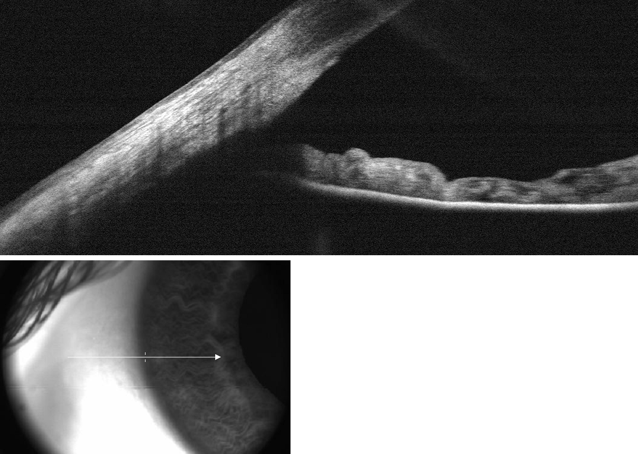
Why the Lower Lid Is Uniquely Vulnerable
The lower lid has no bony shelf below it. It stays pressed against the globe through four overlapping support mechanisms, and blepharoplasty stresses all of them at once.
- Lateral canthal tendon. The outer corner of the eyelid is anchored to the inner surface of the orbital rim by the lateral canthal tendon. This tendon provides primary horizontal tension. When it is lax, the lid bows outward regardless of what else is holding it.
- Orbicularis oculi muscle. This circular muscle encircles the eye and functions as a dynamic sling, pulling the lid inward and upward with every blink. Weakened or disrupted orbicularis - which happens more with the transcutaneous (external-skin) approach than with the transconjunctival approach - removes this active support from the system.
- Lower lid retractors. The capsulopalpebral fascia and inferior tarsal muscle form the posterior lamella support, governing how far the lid descends with downward gaze. Disruption during fat removal can reduce the vertical support from behind.
- Cheek and midface support. The malar fat pad and its ligamentous attachments to the orbital rim provide the foundation the lower lid rests on. As cheek tissue descends with age, that foundation drops and can pull the lid with it - surgery done without addressing cheek descent may worsen an already-falling lid.
Remove even a modest amount of tissue from a system under this much tension and the balance can shift. This is not a design flaw in the surgery; it is the anatomy the surgeon must account for from the first planning conversation.
The Three Ways the Lid Pulls Down
Skin shortage from over-resection
Excessive skin excision is cited in peer-reviewed literature as the single most common preventable cause of post-blepharoplasty ectropion. The anterior lamella has a fixed vertical length. Remove too much and the remaining tissue cannot bridge the distance from the lid margin to the cheek. The lid is pulled downward by geometry. "Conservative skin removal" is not vague advice - surgeons who pinch-test the skin and measure carefully are applying a quantifiable standard, not a personal preference.
Horizontal laxity unmasked by surgery
Some patients arrive with loose horizontal lid tone that causes no symptoms because the intact tissue compensates. Remove anterior lamella support and that compensated laxity becomes structural weakness. The lid that held its position for decades now lacks the backing tissue. This category of retraction is most likely to appear early - within the first weeks - before significant scarring has developed.
Scar contracture over time
The third failure mode is the slowest and the most deceptive. Initial healing looks acceptable, but scar tissue in the anterior lamella matures and shortens over weeks and months. Patients who look fine at the two-week visit may develop progressive lid lowering through week six, week twelve, and beyond. This is cicatricial retraction. It typically peaks around three to four months and then plateaus as the scar fully matures - which is one of the reasons surgeons ask patients to wait before pursuing surgical repair.

The Preoperative Exam That Separates Safe Candidates from High-Risk Ones
A careful surgeon evaluates lower lid competence before agreeing to a transcutaneous approach. Three clinical tests and one anatomical observation do most of the work.
The snap-back test
The examiner pulls the lower lid gently downward, releases it, and watches what happens. In a lid with healthy orbicularis tone, the lid snaps back to its resting position promptly, without the patient needing to blink to reset it. A lid that drifts back slowly - or that requires a blink - signals orbicularis laxity. That laxity may be invisible in daily life, but it predicts meaningful risk once the muscle is disrupted by a transcutaneous incision.
The distraction test
The examiner pulls the lower lid away from the globe horizontally and assesses how far it moves. Published grading - from mild to moderate to severe laxity - corresponds to increasing degrees of horizontal looseness, with each category defined in the clinical literature (StatPearls NBK576403 provides the full grading scale). Patients with moderate or severe laxity on distraction are candidates for a simultaneous canthopexy or canthoplasty to reinforce horizontal support at the time of the original blepharoplasty, not after the lid has already pulled down.
Vector assessment
Viewed from the side - in a profile photo or a mirror - trace an imaginary vertical line down the front of the cornea. In a favorable or neutral vector anatomy, that line falls behind the inferior orbital rim or aligns with it. In negative vector anatomy, the front of the cornea projects forward beyond the inferior orbital rim. The practical consequence: the lower lid must travel a longer, less-supported path to reach the globe. Any surgery that shortens the anterior lamella in this setting starts from a mechanical disadvantage.
Negative vector is common, not rare. It often accompanies high cheekbones that project forward at cheek level rather than at the orbital rim level. Related risk markers for lower-lid malposition - including canthal laxity and the relative position of the lateral canthus - are part of the examination your surgeon should conduct before planning any skin excision (StatPearls NBK576422 covers canthal support assessment in detail). Surgeons who identify negative vector modify the operative plan: often choosing the transconjunctival approach exclusively, adding structural fat grafting, or declining to excise skin at all.
Existing scleral show
A patient who already shows white below the iris before surgery is in a different category from one who does not. Removing skin from a lid already displaying scleral show can convert mild show to frank ectropion. Many experienced surgeons treat even borderline preoperative scleral show as a contraindication to any skin excision on the anterior lamella, regardless of how much loose skin appears to be present.
The Decision Cascade: Approach, Canthus, and Skin Quantity
Transconjunctival vs. transcutaneous
The incision location is the first and most important decision. In the transcutaneous approach, the incision runs just below the lash line on the outer skin. The anterior lamella is opened, the orbicularis is disturbed, and skin may be excised. In one published series of 200 patients, the transcutaneous approach carried a complication rate approaching 10 percent. The transconjunctival approach - incision hidden inside the conjunctiva, no outer skin cut - eliminates skin-shortage retraction entirely because the anterior lamella is never opened or shortened.
| Factor | Transcutaneous | Transconjunctival |
|---|---|---|
| Skin excision possible | Yes | No (separate step if needed) |
| Risk of anterior lamella shortage | Present | Eliminated |
| Orbicularis disruption | More likely | Minimal |
| Suitable for negative vector anatomy | Caution required | Preferred |
| External scar | Faint lash-line line | None externally |
Canthopexy vs. canthoplasty - and when each applies
Canthopexy supports the lateral canthus with sutures without cutting the tendon. It is a relatively brief addition to a lower blepharoplasty, using one or two sutures to reinforce the outer corner without altering the tendon anatomy itself. Canthoplasty goes further: the tendon is severed and reattached at a better-supported position. This is reserved for patients with moderate to severe preexisting laxity. The decision between the two - and whether to perform either - comes directly from the snap-back and distraction test findings. Surgeons who skip those tests have no clinical basis for making the call either way.
After Surgery: What Normal Looks Like and When to Call
Some degree of lower-lid lowering in the first week after surgery is expected. Surgical swelling thickens the tissues and can temporarily hold the lid lower than its final position. Chemosis - swelling of the conjunctiva - can push the lid outward for several days. The question is whether what you are seeing is transient fluid or structural displacement.
- Day 3: Significant swelling is normal. The lower lid may appear lower than it did preoperatively, the skin may feel tight, and mild chemosis is common. This appearance alone does not indicate ectropion.
- Week 2: Swelling should be visibly subsiding. If the lid margin is clearly rolling outward - if you can see the inner pink surface of the lid - or if the eye cannot fully close, contact the surgeon rather than waiting for the next scheduled visit.
- Week 6: The contour of the lid is becoming clearer. Mild lid lowering may still reflect active scar maturation. Persistent scleral show that was not present before surgery, or a visible rolled lid margin, should be photographed and discussed directly at this appointment.
The 6-12 Month Conservative Phase: What It Actually Involves
Most surgeons advise waiting six to twelve months before offering surgical ectropion correction after lower blepharoplasty. This period is not passive delay. Scar tissue continues to remodel for up to a year, and operating on active scar carries a real risk of recurrence and worsening. The conservative phase is also genuinely therapeutic in its own right.
- Lubrication. Preservative-free artificial tears during the day and lubricating gel or ointment at night protect the corneal surface while healing continues. If the lid does not close fully during sleep, ocular ointment is not optional - corneal exposure without lubrication causes abrasion over hours.
- Lid massage. Upward massage of the lower lid - pressing gently from the cheek toward the eye - applies controlled mechanical force against the contracting scar. The technique involves placing a clean fingertip on the lower lid skin and stroking upward, holding gentle pressure at the top of the stroke for a few seconds before releasing. Done consistently over months, it can meaningfully counteract scar shortening. Your surgeon should demonstrate the correct direction and pressure in person - downward massage will worsen retraction, not improve it.
- Taping. Surgical tape applied to the lower lid at night, pulling the lid upward toward the lashes, maintains gentle stretch during the hours when the eye is closed and massage is not being performed. Micropore or paper tape is typically used; test a small patch first for skin sensitivity.
- Steroid injections. For active hypertrophic scar tissue, a series of low-dose steroid injections into the scar band can soften the tissue and slow contracture. This is a clinical decision based on the appearance of the scar, not a universal step in conservative management.
A 2024 clinical study (PMC11626732) evaluated hyaluronic acid filler injected into the preseptal space as a non-surgical option for mild cicatricial lower-lid ectropion. The mechanism is volumizing and stretching the contracted anterior lamella from within - providing mechanical stretch rather than external pressure. For patients with mild cicatricial changes who want to avoid or postpone surgical revision, this is an emerging option to raise with an oculoplastic surgeon, not something to pursue from a general aesthetic injector without eyelid surgical training.
Surgical Repair: Matching the Fix to the Failure Mode
The lateral tarsal strip
For ectropion driven by horizontal laxity, the lateral tarsal strip procedure is the primary repair. The outer corner of the lid is opened, the lid margin is stripped of its mucous membrane, and the resulting strip of tarsus - the firm structural plate within the lid - is anchored to the inner surface of the orbital rim at a corrected position. A retrospective analysis of 85 cases found approximately 95 percent success in restoring lid position with this technique (PMC10577831). It is the most commonly performed ectropion repair because it directly addresses underlying horizontal laxity without requiring donor tissue.
Skin grafting when tissue is genuinely missing
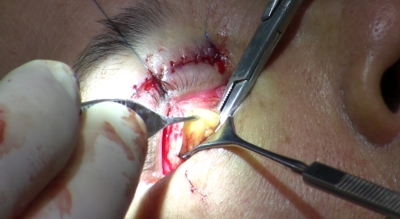
When vertical skin shortage is the cause - as in cicatricial retraction from over-resection - no amount of horizontal tightening restores lid position, because the limiting factor is missing anterior lamella. Repair requires recruiting skin from elsewhere. Upper eyelid skin is generally considered the closest match in thickness, color, and texture and is the most commonly used donor site where sufficient tissue is available. The preauricular area and the retroauricular area directly behind the ear are both well-established alternatives; retroauricular skin is often chosen when upper eyelid donor tissue is insufficient or has already been altered by prior surgery - confirm the best option for your situation with your surgeon. Patients consenting to this repair should understand that there are two recovery wounds: the eyelid graft site and the donor site. The donor site heals well but remains tender for several weeks, requires basic wound care, and leaves a small linear scar that sits in a location typically not visible in normal daily settings. The graft on the eyelid itself is often bolstered or taped for the first week to keep it in contact with the underlying tissue while it takes, and it may look slightly thickened or discolored for several months before softening to a more natural appearance.
Midface elevation
When cheek descent is contributing to lower-lid retraction - pulling the lid down as the midface drops - tightening the lower lid alone may not hold long-term because the foundation beneath it keeps falling. Midface or cheek elevation, through deep-plane or endoscopic approaches, restores the support structure the lower lid depends on. This adds significant complexity and recovery to any revision and is reserved for cases where the anatomy clearly shows descended cheek support as a component of the problem, not a routine addition to tarsal strip repair.
Who Should Perform the Repair
Lower-lid ectropion repair after blepharoplasty is revision surgery on already-altered anatomy. Soft tissue planes are changed, landmarks are obscured by scarring, and the margin for error is narrower than in a primary operation. The skill set required is genuinely specialized.
Oculoplastic surgery - the subspecialty that combines ophthalmology training and reconstructive plastic surgery specifically around the orbit and eyelids - is the most directly applicable background for managing post-blepharoplasty lower-lid complications. Facial plastic surgeons with a concentrated eyelid practice are a second appropriate option.
The question of whether to return to the original surgeon or seek a specialist is one that patients often find awkward. Consider these practical factors:
- If the original surgeon is an oculoplastic specialist with ectropion repair as an active part of their practice, returning is reasonable - they already know your anatomy from the primary operation.
- If the original surgeon is a general plastic or cosmetic surgeon who performs blepharoplasty among a wide variety of procedures, a consultation with a dedicated oculoplastic surgeon is appropriate. This is not an accusation; it is acknowledging that lower-lid revision surgery is a subspecialty skill.
- A surgeon who discourages a second opinion for a complication of this type, or who proposes immediate reoperation before the scar maturation window has closed without a compelling clinical reason, is itself a signal to seek that second opinion.
- Begin documenting with photographs now if you have not already. Date-stamped serial photos taken from the same angle and lighting are the most useful clinical information a revision surgeon receives at a first consultation - more useful than verbal descriptions of how the lid has changed over time.
Frequently Asked Questions
How do I check for negative vector anatomy myself before a consultation?
Stand sideways in front of a mirror in good light and look at your profile. Trace an imaginary vertical line down the front of your eye. If the lower part of your cheekbone projects forward to meet or exceed that line, you have a more favorable vector. If your cheekbone appears to sit behind the front of your eye - recessed relative to the cornea - your lower orbital rim may not be providing enough anterior support, which is the negative vector configuration. A surgeon's assessment is definitive, but this profile check gives a useful starting point for the conversation.
My lower lid looks lower than before surgery but my surgeon says it is swelling - how do I tell the difference?
The distinguishing signs are trend and structure, not absolute position. Swelling-related lid lowering improves week over week; structural displacement does not, or worsens. More specifically: if the lid margin remains in contact with the globe without rolling outward, and the eye closes fully at night, swelling is a plausible explanation. If the pink inner surface of the lid is visible, or you cannot fully close the eye, contact your surgeon the same day rather than waiting for the next appointment.
Can mild ectropion after blepharoplasty resolve on its own?
Mild scleral show from temporary orbicularis weakness can improve as muscle tone recovers over several months. Early cicatricial changes sometimes partially release as the scar softens during its maturation phase. Frank ectropion - where the lid margin rolls outward and the conjunctiva is exposed - very rarely resolves without any intervention. The conservative measures described above (massage, taping, lubrication) work by influencing scar biology, not by recovering tissue that was excised or by repairing tendon laxity, so their effectiveness depends on the underlying cause.
Why do surgeons wait so long before operating to fix ectropion?
Scar tissue is biologically active for up to twelve months after surgery, continuously remodeling and often partially releasing contracture on its own. Operating on immature scar triggers a fresh inflammatory response that can worsen the contracture and significantly raise recurrence rates. The waiting period also allows time to assess whether conservative measures - massage, taping, steroid injections - will resolve the problem without a second operation. Surgeons who enforce this timeline are working with the biology of healing, not delaying care unnecessarily.
If a skin graft is needed, what does recovering from the donor site look like?
Recovery from the skin graft donor site - whether preauricular, retroauricular, or upper eyelid - typically involves tenderness for roughly two to three weeks and a need to keep the area clean during that period. Most patients describe it as noticeable but manageable discomfort rather than significant pain. The donor site heals into a small scar whose visibility depends on its location; preauricular and retroauricular scars sit in natural shadows that are typically not visible in normal daily settings. The graft on the eyelid itself is often bolstered or taped for the first week to keep it in contact with the underlying tissue while it takes, and it may look slightly thickened or discolored for several months before softening to a more natural appearance.
If I developed retraction after my first blepharoplasty, can I have lower-lid surgery again?
Yes, but the risk profile is higher and the approach must be modified to account for what happened the first time. A surgeon planning revision lower blepharoplasty on someone who developed retraction previously will typically use the transconjunctival approach exclusively, avoid any anterior skin excision, and plan a simultaneous canthopexy or tarsal strip. The underlying anatomy - vector, horizontal laxity, orbicularis tone - needs to be formally reassessed, not assumed to be identical to what was found before the first surgery.
General information only, not medical advice. Individual anatomy and healing vary. See the disclaimer.