Most patients seeking lower eyelid surgery understand the goal - reduce those bags under the eyes. What very few understand is that the surgeon faces a genuine fork in the road before the first incision: take the fat out, or move it somewhere more useful. These are not stylistic variations. They produce fundamentally different outcomes, rest on opposite anatomical logic, and carry different risks and recovery timelines. Understanding which approach applies to your anatomy - and why - is the most important question you can bring to a consultation.
The Bag Is Not Extra Fat - It Is Normal Fat That Has Shifted
The lower eyelid contains orbital fat, the same fat that cushions the eyeball inside the bony socket. This fat is not a cosmetic excess the body miscalculated. It belongs there, surrounding and protecting the eye. The problem is not the fat itself but what is holding it in place - or failing to.
Separating the orbital fat from the surface of the lower lid is a thin fibrous membrane called the orbital septum. In youth, the septum is taut enough to keep the fat behind the orbital rim, creating a smooth transition from eyelid to cheek. With age, the septum weakens through a process called involutional laxity - it simply loses its tension. As resistance drops, normal pressure inside the orbit pushes the fat forward. The fat does not grow. It herniates - it bulges through the weakened membrane the way a tire pushes through a cracked sidewall.
That bulge is what appears as an under-eye bag. This is why fat repositioning makes anatomical sense: if the problem is fat that has shifted from where it should be, moving it to where it is actually needed is a more logical correction than removing it from the body entirely.
Anatomy of the Three Fat Compartments
The fat behind the lower eyelid is not a single uniform mass. It is organized into three distinct compartments - medial, central, and lateral - each with its own characteristics and surgical implications.
- Central compartment: The largest of the three and the most likely source of the classic rounded bag. When patients point to a bulge at the center of the lower lid, they are almost always pointing to central fat herniation. This pad responds well to both excision and repositioning.
- Medial compartment: Sits closest to the nose. The fat here is distinctly paler and denser than in the other compartments, enclosed in a thicker capsule, and positioned more deeply in the orbit. Its character is different enough that careful tissue identification is essential before any work proceeds in this area. Note that the lacrimal gland - the tear-producing gland, located in the superolateral orbit - is a structure surgeons must identify and protect primarily when accessing the lateral fat compartment, where it can be inadvertently encountered; it should not be mistaken for fat in any compartment, and any manipulation risks affecting tear production.
- Lateral compartment: The smallest of the three. It tends to herniate less prominently and is typically the least symptomatic, though it still requires assessment during any lower blepharoplasty.
Between the medial and central compartments runs the inferior oblique muscle, one of the six muscles that control eye movement. Its actions include extorsion (outward rotation of the eye), elevation, and abduction. The arcuate expansion of this same muscle also defines the boundary between the central and lateral compartments. In practical terms, the inferior oblique is the most consequential anatomical landmark in lower eyelid surgery.
The Arcus Marginalis - The Ledge That Matters Most
Running along the lower edge of the orbital rim is a fibrous band called the arcus marginalis. This is the point where the orbital septum inserts into the periosteum - the thin connective tissue covering the bone of the orbital rim. Structurally, the arcus marginalis is a ledge. It marks the exact boundary between the eyelid above the rim and the cheek below it.
Just below that ledge, in many patients, lies a hollow groove running inward from the inner corner of the eye toward the outer cheek. This is the tear trough, or nasojugal groove. In youth, the eyelid-to-cheek transition is smooth. As the midface loses volume with age and the cheek descends, the tear trough becomes a visible depression that casts a shadow and reads as fatigue or premature aging. Critically, this midface volume loss also makes the fat herniation above the rim look worse than it actually is - a bulge sitting above a deep groove appears larger and more dramatic than the same bulge sitting above a smooth cheek.
This is the key mechanical insight: fat repositioning addresses both problems at once. The herniated fat above the rim and the hollow groove below it are separated by the arcus marginalis. When a surgeon releases that fibrous band, the fat from above the rim can be brought across and draped into the tear trough beneath. The bulge above becomes the filler below. No other single maneuver accomplishes the same correction.
Why Surgeons Used to Remove the Fat - and Why That Created a New Problem
From the earliest lower blepharoplasties through most of the 1980s, direct fat excision was the standard technique. The orbital septum was opened, the fat was pulled forward, and the excess was cut away. The rationale was simple: the fat bulges, so remove what bulges.
Fat transposition was first described by Loeb in 1981, but it was Sam Hamra's landmark 1995 paper in Plastic and Reconstructive Surgery - combining arcus marginalis release with fat repositioning - that brought the concept into mainstream practice. The shift in thinking was not immediate.
What drove the change was a pattern that aggressive excision reliably produced. Removing large volumes of orbital fat left patients with a hollowed, skeletonized appearance around the eye socket. The orbital rim became visible as a hard ridge under thin skin, and the under-eye area looked sunken rather than refreshed. That effect worsens over time as surrounding tissue continues to lose volume, making the hollowness more pronounced rather than less. A patient who looked adequately improved at six months could look drawn and unnatural at five years.
Removing fat eliminates the bag but leaves nothing to address the groove below it - and as the face ages, that groove only deepens.
The deeper problem is reversibility. A hollow orbital rim caused by over-resection is genuinely difficult to correct. Fat grafting can partially restore volume, but the texture and predictability of grafted fat differs from the original tissue. Most surgeons would rather move fat than explain why it needs to be brought back from somewhere else years later.
How Fat Repositioning Actually Works, Step by Step
Understanding the sequence makes the surgical logic concrete rather than abstract.
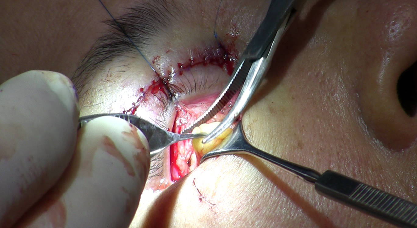
- Incision placement. For transconjunctival repositioning, the incision goes inside the lower lid - through the conjunctiva, the moist membrane lining the inner surface. No external cut, no visible scar on the skin.
- Opening the orbital septum. The surgeon cuts through the septum to access the fat compartments. The inferior oblique muscle is identified and protected before any work proceeds in the space between the medial and central pads.
- Fat mobilization. Rather than pulling the fat out to excise it, the surgeon keeps the fat attached to its vascular supply and gently frees it as a pedicle - a tissue flap that stays connected and therefore maintains blood flow through the repositioning.
- Releasing the arcus marginalis. The surgeon cuts through the fibrous insertion band at the orbital rim. Before this step, the fat cannot travel below the rim. After it, the path is open.
- Draping the fat into the tear trough. The mobilized fat pad is brought over and below the orbital rim and positioned into the depression of the tear trough. The surgeon judges how much volume to place and how to distribute it along the groove.
- Suture fixation. The fat is sutured into the subperiosteal or supraperiosteal plane - anchored against or just above the bone surface - so it holds its new position while the surrounding tissue heals and incorporates it.
Published outcomes data on the transconjunctival approach is encouraging. One reported series found that a large majority of patients achieved substantial elimination of both fat bulges and tear-trough deformity without any skin excision - a result that would be anatomically impossible with excision-only technique, which removes the very tissue used to fill the groove. Specific outcome rates vary by series and patient selection; ask your surgeon what their own results look like for your anatomy.
When Fat Removal Is Still Correct
The shift toward repositioning is not absolute. There are anatomical situations where excision remains the right decision, or at least part of it.
- True fat volume excess. In some patients, the total volume of herniated fat genuinely exceeds what the tear trough depression can hold. Transposing all of it would produce an overfilled groove rather than a smooth correction; excess must be removed.
- Very prominent herniation. When a fat pad herniates severely and extends well beyond the orbital rim, the anatomy may not lend itself to clean repositioning without leaving too much bulk below the rim.
- Minimal or absent tear trough. Repositioning works because there is a hollow to fill. A patient without a significant tear trough deformity has less to gain from the maneuver, and conservative excision may produce a cleaner result with simpler recovery.
The critical distinction is between selective, conservative excision of genuinely excess fat and the older philosophy of wholesale removal. The modern standard is to excise only what cannot usefully be repositioned - and to reposition what can.
The Hybrid Approach: Reposition Some, Remove Some
Most patients presenting for lower blepharoplasty do not fall neatly at either extreme. The majority fall in the middle - enough fat to need some reduction, enough tear trough to benefit from some transposition. For these patients, the hybrid approach is now the predominant technique among experienced surgeons.
A study published in Plastic and Reconstructive Surgery (indexed on PubMed, 2024-2025) examined the hybrid technique in the transcutaneous approach: releasing the arcus marginalis, transposing the septum-fat flap over the orbital rim, and selectively resecting fat from the deeper layer. The study documented strong outcomes with the combined approach, with 91.6% of patients achieving complete elimination of fat bulges and tear-trough deformity. The logic reflects what anatomy suggests - use the superficial fat to fill the groove, remove what would otherwise create overfill.
In practice, a hybrid procedure works like this: the surgeon opens the fat compartments, assesses total volume, repositions the anterior portion of the central and lateral fat into the tear trough, then trims the deeper, more posterior fat that would otherwise remain as excess. The medial fat pad, with its thicker capsule and deeper position - and its proximity to important structures at the medial orbital margin - is often approached more conservatively, with careful identification before any tissue is moved or removed.
Incision Choice: Transconjunctival vs. Transcutaneous and How It Relates to Fat Management
| Factor | Transconjunctival | Transcutaneous |
|---|---|---|
| Incision location | Inside lower lid, no skin cut | Skin, just below the lash line |
| External scar | None | Fine line at lash margin |
| Skin removal | Not possible via this route alone | Concurrent skin excision possible |
| Retraction / ectropion risk | Lower | Higher due to external tissue disruption |
| Fat repositioning | Preferred route when skin laxity is minimal | Allows repositioning and skin work together |
| Best candidate | Fat herniation with minimal skin laxity | Fat herniation with significant skin excess |
The incision choice is not separate from the fat management decision - it constrains what is possible. A transconjunctival approach cannot address loose lower lid skin. When a patient has significant skin laxity in addition to fat herniation, a transcutaneous approach or a combined approach becomes necessary: transconjunctival fat repositioning paired with a separate small skin excision or skin-tightening treatment at the lower lid.
The transcutaneous route carries a meaningfully higher risk of lower eyelid retraction and ectropion - a condition where the lower lid pulls downward and away from the eye. This occurs because the external incision disrupts tissue planes that support the lid position. In experienced hands with appropriate patient selection, the risk is manageable, but it is not trivial and should factor into any discussion of technique when skin laxity is borderline.
What to Ask Your Surgeon Before You Book
These six questions go beyond the standard consultation checklist. They are designed to surface the specific anatomical thinking that separates surgeons with genuine lower blepharoplasty depth from those applying a single technique to every face.
- "Do you reposition the fat, remove it, or use a combination - and how do you decide for a given patient?" The answer should be anatomy-based. A surgeon who always does one approach regardless of anatomy should be able to articulate exactly why your anatomy fits that approach.
- "How do you assess tear trough depth, and does my anatomy suggest repositioning would benefit it?" If the surgeon has not evaluated your tear trough independently from your fat herniation, the fat plan may be incomplete.
- "If I also have skin laxity, how does that change your incision choice and your approach to the fat?" Skin laxity and fat herniation require coordinated decisions. The answer reveals whether the surgeon plans these together or treats them as separate problems.
- "How do you identify and protect the inferior oblique muscle during fat compartment work?" A surgeon who can describe the muscle's position, its role as a compartment divider, and their specific technique for protecting it has internalized the anatomy in a meaningful way.
- "How do you identify and protect the lacrimal gland and other key structures when working near the lateral and medial fat compartments?" This question surfaces the surgeon's awareness of the structures at risk in each part of the orbit and how they distinguish fat from other tissue before removing or moving anything.
- "What should I realistically expect at four weeks versus three months after surgery?" The correct answer acknowledges that repositioned fat swells longer and that the tear trough result takes closer to three months to stabilize. A surgeon who promises a complete result at one month has not adequately prepared patients for this recovery.
Frequently Asked Questions
Is fat repositioning more effective than fat removal for under-eye bags?
For most patients - particularly those with a visible tear trough in addition to fat herniation - repositioning produces better long-term outcomes because it addresses both the bulge above the orbital rim and the hollow below it in a single step. Simple fat removal eliminates the bulge but leaves or worsens the groove. That said, the right technique depends on each patient's fat volume, tear trough depth, and skin condition, which is why preoperative assessment matters more than a blanket technique preference.
Will I have a visible scar after lower eyelid fat repositioning?
If the transconjunctival route is used - incision placed inside the lower lid through the conjunctiva - there is no external scar. The transcutaneous approach places a fine incision just below the lash line, which heals to a nearly imperceptible line in most patients, but it is an external cut. Which route is appropriate depends primarily on whether you also need skin excision at the lower lid.
Can the hollowness caused by previous fat removal be corrected?
Yes, though correction is more involved than a well-planned primary procedure. Options include fat grafting to the orbital zone, injectable fillers placed carefully in the periorbital area, or in some cases secondary repositioning of any remaining fat. Results are less predictable than primary surgery. This is one of the main reasons current surgical thinking favors conservative fat handling from the outset rather than aggressive removal that may later need reversal.
Why does swelling last longer after fat repositioning than after excision?
The repositioned fat is transferred as living tissue that must grow new blood vessels in its new location - a biological process called revascularization. Until that process completes, the tissue retains more fluid and swells more persistently. Simple excision removes tissue entirely, so there is less living tissue remaining to swell. Patients undergoing repositioning should plan on the tear trough area taking two to three months before the result is fully visible.
Can fat repositioning be done at the same time as treatment for skin laxity?
Yes. When skin laxity is significant, surgeons use either the transcutaneous approach - which allows skin excision from the same incision - or a combined approach: transconjunctival fat repositioning paired with a separate skin excision or skin-tightening step. The combination addresses fat herniation, tear trough volume, and excess skin without requiring a trade-off between them. The planning for each element should happen together, not as afterthoughts to one another.
How do I know if my tear trough is deep enough to benefit from fat repositioning?
A groove visible under your eyes - particularly one that casts a shadow and reads as fatigue even when you are rested - suggests anatomy where repositioning could be beneficial. The assessment requires an in-person examination by a surgeon who evaluates the tear trough separately from the fat herniation and considers how they interact. Photographs taken in different lighting conditions are often part of a thorough preoperative review because the depth of the groove and the severity of the herniation can look very different under different light.
General information only, not medical advice. Individual anatomy and healing vary. See the disclaimer.