When facial nerve palsy strikes, the eyelid stops moving. That sounds like a cosmetic problem. It is not. Within hours of nerve failure, the exposed cornea begins to dry, the tear film collapses, and damage that follows can become permanent. This guide covers every stage of that process - from the anatomy of a blink to the surgical choice between a gold weight and a platinum one - for the full range of patients affected: not only those recovering from Bell's palsy, but those who had acoustic neuroma surgery, parotid procedures, or other head and neck operations that placed the facial nerve at risk.
How Facial Nerve Palsy Disables the Eyelid
Two muscles, two separate nerve supplies
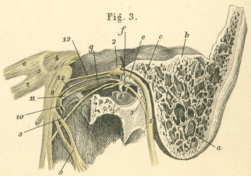
The eyelid is controlled by two opposing muscles. The levator palpebrae superioris opens the upper lid; it is driven by the third cranial nerve and is unaffected in facial palsy. The orbicularis oculi closes both lids in a smooth circular sweep; it is driven entirely by the seventh cranial nerve, the facial nerve, through its zygomatic and buccal branches. When the facial nerve fails, the orbicularis stops working but the levator keeps pulling the lid upward. The result is an eye held open by an unopposed muscle - wide, unblinking, unprotected.
This is not simply a failure to blink consciously. Every reflex blink - the automatic spread of tears across the corneal surface that happens every few seconds - requires orbicularis contraction. Without it, the tear film stops refreshing. The mucin layer that anchors tears to the cornea begins to break down within the first hour of exposure. The epithelial cells on the corneal surface depend on constant hydration and the mechanical wipe of a closing lid; deprived of both, they dry and begin to die. The conjunctiva reddens. By the end of the first day without protection, the cornea can already show punctate erosions visible under a slit lamp.

Causes Beyond Bell's Palsy
Bell's palsy - sudden-onset facial paralysis of presumed viral origin - affects roughly 15 to 40 people per 100,000 each year, with a lifetime risk in the range of 1 in 60. It is the single most common cause of peripheral facial nerve palsy and therefore the most common cause of paralytic lagophthalmos. But it is far from the only cause, and patients who developed palsy through other routes often find that most published patient information simply does not describe their situation.
- Acoustic neuroma surgery (vestibular schwannoma removal): Published rates of delayed postoperative facial palsy following acoustic neuroma excision range from about 14.5% to nearly 30%, depending on tumor size and surgical approach. The nerve may be stretched, compressed, or deliberately sacrificed to achieve full tumor removal. Patients facing this operation should speak with an oculoplastic surgeon before the procedure - not afterward.
- Parotid gland tumor excision: The facial nerve runs directly through the parotid gland, dividing it into superficial and deep lobes. Any tumor that abuts the nerve creates a risk of stretch or transection during removal. The larger or more adherent the tumor, the higher that risk.
- Ramsay Hunt syndrome (herpes zoster oticus): Reactivation of the varicella-zoster virus in the geniculate ganglion produces a painful ear rash alongside facial palsy. It tends to cause more complete and longer-lasting paralysis than Bell's palsy, raising corneal risk proportionally.
- Lyme disease: Facial nerve palsy is one of the most common neurological complications of Lyme disease and can occur bilaterally - affecting both eyes at once - which creates compounded management challenges.
- Brainstem stroke: A stroke affecting the pons can disrupt the facial nerve nucleus, producing lower motor neuron-pattern palsy indistinguishable from peripheral causes in its effect on lid closure.
- Other head and neck surgery: Neck dissection, mastoidectomy, and temporal bone resection can injure extracranial facial nerve branches. Lagophthalmos sometimes appears as a postoperative complication that neither surgeon nor patient anticipated fully.
The Corneal Danger Cascade
Corneal injury in facial palsy does not happen all at once. It follows a predictable sequence, and understanding each step clarifies why early intervention is not optional.
- Exposure: The lid stays open, the tear film evaporates faster than it is replenished, and the cornea is left unprotected against air, light, and foreign particles.
- Exposure keratopathy: The corneal epithelium dries and begins to stain with fluorescein dye under a slit lamp - punctate keratopathy, thousands of tiny surface cell disruptions visible as bright green dots.
- Corneal erosion: Patches of epithelium detach, leaving raw painful areas. These erosions heal slowly without lid protection because every sleep period is another hours-long exposure event.
- Corneal ulceration: Bacteria colonize the damaged epithelium. Infectious ulcers develop and can progress from mild to vision-threatening within 24 to 48 hours.
- Scarring: Even after an ulcer heals, the scar tissue sits permanently in the visual axis. If the scar is central, the resulting blur cannot be corrected with glasses. The only treatment at that stage is corneal transplantation.
Bell's phenomenon: the hidden variable
Approximately 75% of people have Bell's phenomenon - the automatic upward and outward rotation of the eye that occurs when the lids attempt to close. Even in facial palsy, when the lids barely move, this reflex still pulls the cornea up and partially behind the upper lid on each blink attempt, offering meaningful protection against overnight exposure. The remaining 25% of people lack this reflex entirely; their corneas sit stationary and fully exposed through every sleep cycle. That group faces disproportionately higher risk and needs more aggressive early management than someone with the same degree of lid paralysis who does have the reflex. Surprisingly, this distinction appears on almost no patient-facing sources - yet it may be the single factor that tips a patient from manageable exposure toward corneal ulceration.
Double jeopardy: when corneal sensation is also lost
About 10% of Bell's palsy patients have reduced or absent corneal sensation in addition to lid paralysis, caused by simultaneous involvement of the trigeminal nerve. The corneal reflex - the involuntary blink triggered by touching the cornea - depends on the trigeminal nerve sensing contact and the facial nerve executing the blink. When both fail, the condition is called neurotrophic keratopathy. Without sensation, patients cannot feel pain when their cornea is damaged, so the warning system that would otherwise drive them to seek help is missing. For patients who had skull-base surgery - including acoustic neuroma removal - this combination should be specifically checked at the first postoperative visit.
Assessing Risk at the First Visit
The first ophthalmology or oculoplastic visit after facial palsy involves several specific tests that together define how much danger the cornea is in and how urgently treatment must escalate.
- House-Brackmann grading: This six-level scale - Grade I is normal function; Grade VI is complete paralysis with no movement whatsoever - gives a standardized measure of palsy severity. At Grade III, patients can still achieve effortful but complete eye closure, so the cornea retains some protection. At Grade IV and above, complete closure is gone and corneal protection becomes an urgent clinical priority rather than a preventive one.
- Bell's phenomenon test: The examiner asks the patient to close the eye firmly while holding the lid open with a fingertip. The eye's movement - or absence of it - reveals whether the protective reflex is working. A flat, stationary cornea during attempted closure signals higher risk and shifts management accordingly.
- Corneal sensation testing: A wisp of sterile cotton or a dedicated aesthesiometer is brushed lightly across the corneal surface. Normal sensation produces a reflexive blink and the patient reports feeling the contact. Reduced or absent response places the patient in the neurotrophic keratopathy risk category, requiring more intensive lubrication and earlier surgical planning.
- Slit-lamp examination: Fluorescein dye dropped into the eye stains any area of epithelial damage bright green under blue-wavelength light. Punctate staining across the lower half of the cornea is the classic early sign of exposure keratopathy. A larger confluent staining pattern or visible epithelial defect changes the management urgency entirely.
Medical and Conservative Management
For most patients, treatment starts without surgery. The goal is to replicate what the lid normally does: keep the corneal surface wet, reduce evaporation, and protect against overnight exposure when lubrication wears off fastest.
Preservative-free artificial tears are the foundation. Preservatives in standard tear drops accumulate on the corneal surface with frequent dosing and become toxic to the epithelial cells already under stress from exposure. Drops are used every one to two hours while awake, increasing to every hour if the cornea already shows staining. At night, a thick lubricating ointment is applied; it blurs vision so it is not practical during waking hours, but it provides hours of protection through sleep.
Moisture chamber goggles - sealed eyewear with a foam gasket that traps a humid microenvironment around the eye - add another protective layer, particularly during sleep and in environments with low humidity or airflow such as wind, air conditioning, or electric fans. Many patients find that taping the lid closed with paper tape at bedtime provides enough seal when goggles are not tolerated, though some instruction is needed to avoid inadvertently dragging the lid against the eye during removal.
These measures are genuinely sufficient for mild palsy - Grade II or III - when the lid can be coaxed to near-closure with effort and Bell's phenomenon is present. For Grade IV and above, or for anyone lacking Bell's phenomenon whose cornea already shows punctate staining, lubricants alone are buying days rather than solving the problem. That is the signal to advance to the next tier of management without delay.
Botulinum Toxin Ptosis as a Bridge
When a clinician wants to protect the cornea now but does not yet know whether the facial nerve will recover - which is the situation with fresh Bell's palsy or with post-surgical palsy before the nerve's fate is clear - a temporary measure can bridge the gap without committing to a permanent implant.
Botulinum toxin type A is injected into the levator palpebrae superioris, the muscle that opens the upper lid. A carefully measured dose paralyzes the levator and allows the lid to drift downward under gravity. The resulting drooped lid - called a pharmacological or chemical ptosis - covers the cornea and restores most of the protection that the failed orbicularis can no longer provide. Onset takes four to seven days. The duration is roughly six to twelve weeks. When it wears off, the levator recovers completely and the lid reopens as before.
If the facial nerve has recovered enough during that window, no further treatment may be needed. If not, the injection can be repeated, an implant can be planned, or both. Patients find the appearance - a noticeably drooping lid - uncomfortable socially, but most accept it once they understand it is fully temporary and that the alternative is progressive corneal scarring. The term chemical tarsorrhaphy is used by analogy with the surgical procedure that stitches the lids together; unlike the surgical version, it leaves no scar and requires no reversal procedure. This option is almost entirely absent from consumer-facing information about facial palsy, despite being well established in oculoplastic practice.
Eyelid Weight Implants: Gold and Platinum
How the surgery works
An eyelid weight implant is a small plate placed between the orbicularis oculi and the tarsal plate of the upper lid through a crease incision that follows the natural skin fold. The implant sits in front of the tarsus and uses gravity to tip the lid downward on attempted closure. When the patient is upright and looking ahead, the weight has minimal visible effect. When the patient relaxes or looks down, gravity pulls the weighted lid toward the globe - exactly the motion needed when the orbicularis cannot do the job.
Before surgery, the correct weight is determined using adhesive trial weights taped to the skin of the upper lid. The patient walks around, looks in a mirror, and blinks while the surgeon observes. Too light and the lid still gaps; too heavy and it droops noticeably in primary gaze. The right weight achieves functional closure without the lid sitting low during ordinary activity.
Gold versus platinum: a comparison
| Feature | Gold | Platinum |
|---|---|---|
| Relative density | Lower - requires a larger implant to reach the same closing force | Higher - same closing force in a smaller, thinner implant (roughly 11% smaller) |
| Conformity to lid curvature | Solid plate; stiffer against the curved tarsus | Often a chain or segmented design; conforms more naturally to the lid |
| Visibility through thin skin | More visible, especially in patients with thin or aged lids | Lower profile; less visible contour through the skin |
| Allergy and extrusion risk | Extrusion documented; gold allergy rare but possible | Lower rate at high-volume centers; platinum allergy extremely rare |
| Reversibility | Fully removable through the same lid crease incision | Fully removable through the same lid crease incision |
| Current preference at specialized centers | Still appropriate; widely available | Preferred material at most high-volume oculoplastic practices |
The difference in density between the two metals means that a platinum implant achieving the same closing force is physically smaller - roughly 11% lighter by volume than its gold counterpart. That size difference matters in the upper lid, where there is very little tissue between the implant and the skin surface. High-volume centers have largely shifted to platinum for most cases because the thinner profile is less detectable and the extrusion rate is lower. Gold remains entirely appropriate and effective, particularly where platinum implants are not available or when the surgeon's experience favors it. The closure rates reported in published literature - complete or near-complete closure in roughly 83 to 92% of cases - apply to both materials.
Lower Lid Paralytic Ectropion and Its Correction
The orbicularis oculi does not only close the upper lid. It runs in a continuous loop around the entire eye, and its lower portion holds the lower lid against the globe. When the facial nerve fails, the lower lid loses this muscle support and begins to sag outward - a condition called paralytic ectropion. The lower lid pulls away from the eye, the punctum (the tear drainage opening at the inner corner) rotates away from the globe and can no longer drain tears properly, and the now-exposed lower conjunctiva thickens from chronic irritation.
This compounds the corneal problem significantly. The lower tear lake - the reservoir of fluid that the lower lid normally keeps in contact with the cornea - drains away from the eye instead of onto it. The gap between the lower lid and the globe creates another entry point for evaporation and debris. Even a well-fitted upper lid weight loses some of its value if the lower lid is not in contact with the globe, because the two lids meet to complete the blink.
The standard corrective procedure is a lateral tarsal strip. The surgeon detaches the outer corner of the lower lid, shortens and tightens the tarsal strip, and re-anchors it to the periosteum at the outer orbital rim. This tightens and elevates the lower lid back against the globe. In more severe cases - particularly when the cheek and midface have descended over time due to chronic orbicularis weakness - a midface suspension procedure lifts the soft tissue of the cheek upward to support the lower lid from below. These procedures are typically performed at the same operative session as upper lid weight placement, addressing both lids at once.
Reversibility and Long-Term Planning
Lid weight implants are not permanent commitments. If the facial nerve recovers, the implant comes out through the same lid crease incision, under local anesthesia, as an outpatient procedure - and the eyelid returns to normal function.
This fact - that the surgery is straightforwardly reversible - changes the decision for many patients who are hesitant to proceed while nerve recovery is still possible. The window in which reversibility matters is real: Bell's palsy recovers partially or fully in the majority of patients within three to six months, and post-surgical palsy can continue improving for a year or more. Waiting out the full recovery period without implant protection, however, risks permanent corneal damage during those months of uncertainty.
The practical bridge-to-implant sequence
The approach at most centers is to use botulinum toxin ptosis in the first six to twelve weeks. If recovery appears on nerve testing or clinical examination, the injection is repeated until the nerve has recovered enough to manage without it. If recovery is absent or incomplete beyond three months - particularly after a surgical nerve injury - a lid weight is placed, with the explicit understanding that it will be removed when the nerve no longer needs the help. Removal is a minor outpatient procedure performed under local anesthesia. The implant is lifted out; no reconstruction is required. Recovery is a matter of days.
When recovery is partial
Partial nerve recovery creates a more nuanced decision. If the lid can close to within 1 to 2 mm and Bell's phenomenon is present, some patients manage with lubricants and no implant. Others - particularly those who lack Bell's phenomenon or who still show corneal staining despite adequate lubrication - benefit from a weight even with partial function. The weight does not prevent whatever residual orbicularis function exists from contributing; it adds to it rather than replacing it, and it can still be removed if the nerve continues to improve.
Permanent tarsorrhaphy as a last resort
Permanent tarsorrhaphy - stitching the lid margins together - is now a treatment of last resort. It narrows the visual field, alters appearance permanently, and prevents adequate examination of the cornea. When it is used, it is limited to the outer portion of the lid margin so the central visual axis stays open. It becomes relevant primarily when a patient cannot tolerate a weight for anatomical reasons, or when corneal damage is so severe and ongoing that even a small remaining gap carries immediate risk. For patients facing planned facial nerve sacrifice during acoustic neuroma removal, preoperative consultation with an oculoplastic surgeon should happen before the day of surgery. Knowing that a lid weight can be placed at or immediately after the same operative session eliminates days of unprotected corneal exposure in the highest-risk period - when the nerve is freshly injured and recovery is weeks away.
Frequently Asked Questions
How long does Bell's palsy last, and when should I be worried about my eye?
Most Bell's palsy patients see the first signs of recovery within three weeks, with the majority reaching significant recovery by three to six months. Concern about the eye is immediate - not dependent on how long the palsy has lasted. If you cannot fully close your eye on the day of diagnosis, begin preservative-free artificial tears that day and see an eye care provider within 24 to 48 hours for a corneal examination, not at a follow-up weeks later.
What is Bell's phenomenon and how do I know if I have it?
Bell's phenomenon is the automatic upward and outward rotation of the eye that happens during an attempted blink or forcible lid closure. About three in four people have it. To get a rough sense: try to close one eye forcibly while holding the lid open with a fingertip - if you see white sclera rather than the iris, the eye has rotated upward and the phenomenon is likely present. Your eye care provider can test this formally and tell you what it means for your corneal risk level.
Can the botulinum toxin injection affect my vision or is it safe to drive?
The drooped lid reduces the upper visual field on the treated side, which some patients notice when driving or on stairs. It does not affect clarity through the open portion of the eye. Most patients adapt within a few days. The effect resolves completely when the injection wears off, typically within two to three months, and no intervention is needed to reverse it.
What is the difference between a gold and a platinum eyelid weight, and should I ask which one my surgeon uses?
Platinum is denser than gold, so a platinum implant achieving the same closing force is physically smaller and thinner, which reduces visibility through thin eyelid skin and lowers the chance of the implant becoming prominent over time. High-volume centers have largely shifted to platinum for these reasons. Yes - asking which material your surgeon uses and why is a reasonable and informative question. A surgeon experienced with both materials should be able to explain their choice for your specific anatomy.
If the facial nerve recovers after I have a lid weight implanted, is removal a major procedure?
Removal is a minor outpatient procedure under local anesthesia through the same lid crease incision used for placement. The implant is lifted out; no reconstruction is needed. Recovery is measured in days, not weeks. This is worth knowing before you agree to or decline a weight - it is designed from the outset to come out cleanly if the nerve recovers, and many patients who initially refuse surgery change their mind once they understand removal is straightforward.
How is paralytic lagophthalmos different from lagophthalmos after blepharoplasty?
Post-blepharoplasty lagophthalmos results from too much skin being removed during cosmetic surgery - a mechanical tissue shortage prevents the lid from reaching the globe. Paralytic lagophthalmos results from muscle failure, with normal or even excess skin present. The corneal danger is similar in both cases, but the causes, prognosis, and surgical solutions differ substantially. Paralytic lagophthalmos generally requires a lid weight or nerve-related intervention; post-blepharoplasty lagophthalmos is more likely to need a skin graft or tissue redistribution procedure to restore adequate lid length.
General information only, not medical advice. Individual anatomy and healing vary. See the disclaimer.