A small raised bump near the eyelid margin, a pale scar-like patch that appeared where no scar should be, a spot that bleeds when rubbed and never fully heals - these are the presentations that bring patients into an oculoplastic office with what turns out to be basal cell carcinoma. BCC of the eyelid is the most common eyelid malignancy by a wide margin, and the eyelid's unique anatomy makes it both a site of elevated cancer risk and one of the most demanding locations in the body to reconstruct after tumor removal. The path from biopsy to a functioning, natural-looking eyelid requires coordinated expertise that differs substantially from removing BCC on the cheek or forehead.
Why the Eyelid Is a Preferred Site for BCC
The lower eyelid receives more UV radiation than almost any other facial surface of comparable size. When sunlight reflects upward off water, sand, or concrete, the lower lid sits directly in the path of that scatter - acting as a reflective basin for upward UV bouncing off horizontal surfaces. The skin there is among the thinnest on the body, and its sparse sebaceous gland density makes consistent sunscreen application difficult. Proximity to the eye discourages most people from applying product close to the lash line, and even when they do, the skin does not hold it the way cheek or forehead skin does.
These factors explain why BCC accounts for approximately 85 to 95 percent of all malignant eyelid tumors. Sebaceous carcinoma and squamous cell carcinoma each account for a small fraction of eyelid malignancies in most Western series - published estimates typically place each subtype in the 1 to 5 percent range individually, though combined figures and exact percentages vary by study population and geographic region. Still, the key point is that the vast majority of eyelid malignancies are BCC, and patients searching for information about eyelid cancer often encounter alarming descriptions of aggressive tumors that bear no resemblance to what most eyelid lesions turn out to be. The lower eyelid is the most common site, responsible for roughly 48 percent of periocular BCCs in some series, followed by the medial canthus at about 29 percent, the upper eyelid at 17 percent, and the lateral canthus at 5 percent - though distribution figures vary across studies.
What Eyelid BCC Looks Like
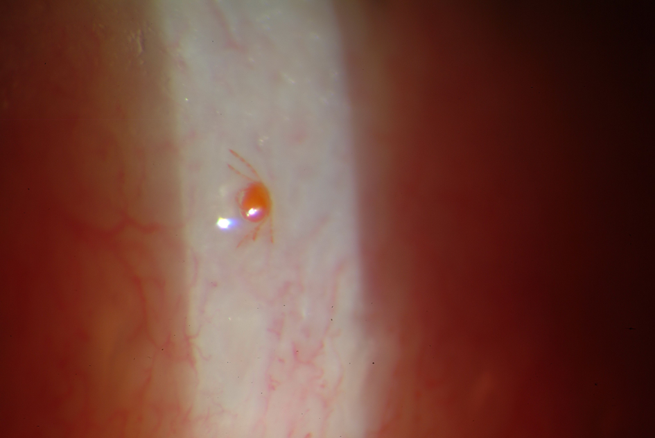
The classic presentation is a nodular BCC: a pearly or translucent nodule with a rolled, slightly raised border, visible surface blood vessels called telangiectasias, and sometimes a central crust or shallow ulceration. In a study of 100 periocular BCC cases, the nodular subtype accounted for approximately 83 percent of presentations. This version looks like a lesion. Patients notice it. They bring it in relatively early.
The morpheaform - or sclerosing - subtype causes diagnostic delay precisely because it does not look like a cancer. It presents as a flat, firm, pale or yellowish plaque with poorly defined edges. The surface looks like scar tissue. Without a history of trauma or surgery to explain it, it is frequently dismissed as a chalazion, blepharoplasty scar, or patch of thickened dry skin. Months and sometimes years pass before a biopsy is done.
The basosquamous or metatypical subtype behaves more like squamous cell carcinoma than typical BCC. It carries a higher rate of perineural invasion and local recurrence, and pathologic identification of this subtype changes both the surgical approach and the surveillance plan.
| Subtype | Clinical appearance | Margin behavior | Recurrence risk |
|---|---|---|---|
| Nodular | Pearly nodule, rolled edges, telangiectasias, possible central ulceration | Visible borders generally correlate with histologic extent | Lower with complete excision |
| Morpheaform | Flat, indurated, scar-like plaque with indistinct edges | Histologic margins extend well beyond visible border | High with standard excision; substantially lower with Mohs |
| Basosquamous | Variable; may resemble an ulcerated or nodular lesion | Higher risk of perineural spread along nerve sheaths | Highest of the three periocular subtypes |
The Biopsy Step
Not every eyelid lesion requires immediate excision. An oculoplastic surgeon evaluating a suspicious lesion will consider how long it has been present, whether it has changed, and its characteristics on close examination. The following features make biopsy the right next step rather than observation:
- A pearly, translucent, or waxy surface with visible surface vessels
- Loss of eyelash follicles along the lid margin where the lesion contacts it
- A lesion that bleeds with minor trauma and fails to heal
- A firm, scar-like patch with no prior trauma or surgery at that site
- Any lesion growing, changing color, or developing ulceration over weeks to months
Biopsy technique matters. For small, well-defined lesions, excisional biopsy - removing the entire lesion - gives the pathologist the most tissue and occasionally achieves clear margins on the first pass. For larger or indistinct lesions, an incisional biopsy establishes subtype before planning definitive surgery. Taking a sample from a morpheaform lesion before committing to a resection approach is appropriate, because the subtype finding directly determines whether Mohs surgery is indicated. The pathology report also identifies perineural invasion, which prompts imaging, and depth of invasion, both of which shape reconstruction planning.

Mohs Surgery and Standard Excision
How margin control differs between the two approaches
Standard wide-local excision removes the visible tumor plus a planned margin of apparently normal tissue, then sends the specimen to pathology. The pathologist examines representative sections - sampling a small fraction of the actual margin surface. For nodular BCC at low-risk sites, this works reasonably well. For eyelid BCC, where morpheaform subtype is common and the eyelid's small size limits the margin width a surgeon can safely take, incomplete margin sampling leaves room for missed tumor. Standard excision carries five-year recurrence rates of approximately 5 to 10 percent for eyelid BCC, and recurrence rates for BCC with positive margins after standard excision can reach 30 to 40 percent - morpheaform BCC is at particular risk of incomplete removal because its histologic extent routinely goes well beyond the visible lesion, which is why Mohs is the preferred approach for this subtype.
Mohs micrographic surgery examines close to 100 percent of the peripheral and deep margin in real time using horizontal frozen sections. If tumor remains, only the involved quadrant is re-excised. The cycle continues until margins are clear. The result is a five-year recurrence rate of approximately 1 percent for primary eyelid BCC - a gap driven entirely by the completeness of margin analysis, not by taking larger margins.
The two-surgeon team and when each approach is chosen
For most eyelid BCCs, Mohs surgery and reconstruction are not performed by the same surgeon. The Mohs surgeon - typically a Mohs-trained dermatologist - handles staged tumor extirpation and margin confirmation. Once the final clear margin is confirmed, the oculoplastic surgeon reconstructs the defect. Patients sometimes assume one surgeon handles everything. In eyelid BCC, the two-specialist model improves both oncologic and functional outcomes. Standard excision with intraoperative frozen section control remains appropriate for certain straightforward cases or where Mohs is not available, but for morpheaform, basosquamous, or medial canthal BCC, Mohs is the standard of care.
The Two-Lamella Problem
The eyelid has a structural organization unlike any other facial surface. It is built in two distinct layers - lamellae - each serving a critical function:
- Anterior lamella: skin and the orbicularis oculi muscle that powers blinking. This is the outer, visible layer.
- Posterior lamella: the tarsal plate (a rigid fibrous structure that gives the lid its shape and stiffness) and the conjunctiva (the mucous membrane that contacts the eye surface with every blink).
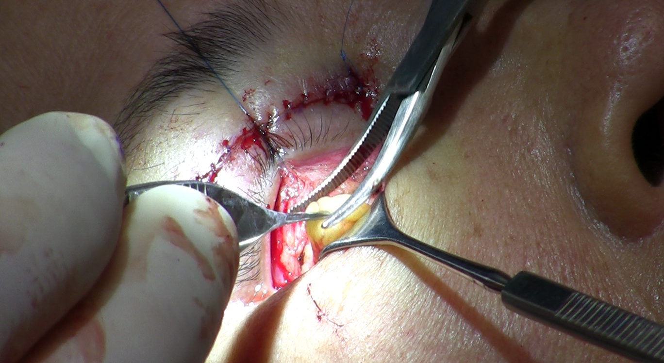
When tumor excision removes tissue from the eyelid margin - the free edge where lashes grow - reconstruction must restore both lamellae. Replacing only the skin layer leaves the lid without structural support. The result is lid retraction, outward turning of the lid edge, and chronic corneal exposure. A cornea exposed to air and friction without adequate lid coverage develops surface damage that can progress to permanent vision loss.
This is what separates an oculoplastic repair from a skin flap alone. A surgeon closing a cheek defect replaces skin and subcutaneous tissue - one functional layer. An oculoplastic surgeon closing an eyelid margin defect rebuilds a two-layer structure that must move smoothly across a sensitive optical surface tens of thousands of times each day. The tarsal plate cannot be replaced by skin. The conjunctival surface cannot be replaced by muscle flap tissue. Specific donor tissues and flap designs are required, and the surgeon must understand orbital anatomy well enough to protect the globe throughout the repair.
The Reconstruction Ladder
Defect size drives reconstruction choice. The principle is to use the simplest approach that safely closes the defect without distorting the eyelid or exposing the cornea.
- Small defects involving up to roughly a quarter of the horizontal eyelid margin can often be closed directly, with layered suture repair of both lamellae.
- Larger defects in the 30 to 50 percent range typically require lateral canthotomy and cantholysis - releasing the lateral canthal tendon to mobilize the remaining lid tissue and allow tension-free closure.
- Semicircular (Tenzel) flap: a crescent-shaped rotation flap from the lateral canthus used when cantholysis alone is not enough to bridge the defect.
- Hughes tarsoconjunctival flap: the standard reconstruction for lower lid defects involving more than half the lid margin, borrowing posterior lamella tissue from the upper eyelid.
- Mustarde cheek rotation flap: a large rotation flap from the cheek used when the defect is extensive enough that local eyelid tissue is insufficient.
The Hughes Flap in Plain Language
The Hughes tarsoconjunctival flap is a two-stage procedure. Patients preparing for large lower eyelid reconstruction deserve a clear account of what each stage involves.
- Stage one - flap creation and inset: Working under the upper eyelid, the surgeon harvests a segment of tarsal plate and conjunctiva from the upper lid's posterior lamella, leaving enough tarsus intact above so the upper lid retains its shape and function. This harvested strip is advanced down to fill the posterior lamella defect in the lower lid. The anterior lamella is rebuilt using a skin graft or local advancement flap from the cheek. At the end of stage one, the upper and lower eyelids are connected by the flap bridge, and the eye is covered to protect it.
- The waiting period: The flap must develop an independent blood supply from the lower lid recipient bed before it can be separated from the upper lid pedicle. The interval varies by surgeon preference and individual healing - ranging from as little as 2 weeks in current practice to 6 weeks or more in more conservative protocols; confirm the specific timeline with your surgical team. During this time, the operated eye is not available for vision. Patients manage daily life with one functional eye - this affects depth perception, driving, and a range of occupational tasks. This is the part of reconstruction that patient-facing resources almost never describe, leaving people unprepared. Arranging workplace accommodations, transportation support, and help at home before stage one is not optional - it is planning the recovery.
- Stage two - flap division: Once vascularization is confirmed, a second procedure divides the tissue bridge connecting the upper lid to the lower lid flap. Excess tissue is trimmed, both lids are sutured into their final positions, and the eye opens. Most patients achieve functional and cosmetically acceptable results after division, though occasional refinement procedures are needed for symmetry or lid position adjustment.
High-Risk Locations and Orbital Invasion
Why medial canthal BCC is in a separate category
BCC at the medial canthus - the inner corner of the eye where the tear drainage system sits - carries the highest local recurrence risk of any periocular location. The anatomy explains why. Tissue planes in the medial canthus allow tumor cells to track subcutaneously toward the lacrimal sac, the nasolacrimal duct, and the orbit, often before any surface change is visible. A lesion that appears small at the skin level may have microscopic extensions reaching the lacrimal drainage system or orbital periosteum. Achieving complete margin clearance here requires more Mohs stages, careful assessment of deep margins near the lacrimal apparatus, and in some cases a dacryocystorhinostomy to restore tear drainage if the nasolacrimal duct is involved.
When imaging is ordered and what it changes
For most small, primary eyelid BCCs, imaging is not part of the workup. That changes when clinical findings suggest deeper invasion. An oculoplastic or orbital surgeon will order MRI or CT when any of the following are present:
- Restricted eye movement or double vision
- Proptosis - forward displacement of the globe
- A lesion that feels fixed or adherent to the orbital rim on palpation
- Numbness in the periorbital skin, suggesting perineural spread
- Recurrent BCC with a history of incomplete excision at the medial canthus
Orbital invasion occurs in approximately 1 to 4 percent of neglected or recurrent periocular BCCs. When involvement is limited to orbital fat or periosteum, globe-sparing surgery with clear margins is sometimes achievable. When tumor has infiltrated the orbit more extensively, orbital exenteration - removal of all orbital contents - becomes the oncologically necessary choice. Patients who encounter descriptions of exenteration without context understandably find them alarming. The critical context: early-stage, primary BCC almost never reaches this point. It is a consequence of long-neglected or multiply recurrent disease, not an outcome of routine diagnosis and timely treatment.
Prognosis and Surveillance After Treatment
The primary risk for a patient who has had periocular BCC is not metastasis - it is local recurrence and new primary BCCs elsewhere on sun-exposed skin, which is why lifelong dermatologic surveillance is a meaningful part of long-term care, not an abundance of caution.
BCC metastasizes at a rate estimated well under one percent over a lifetime. Systemic disease is not the main concern in periocular BCC. Surveillance goals are detecting local recurrence early - before a second tumor requires a more complex second reconstruction - and identifying new primary BCCs at other sites. Most surgeons schedule return visits at 3 to 6 month intervals for the first one to two years, then annually. Recurrence after Mohs surgery tends to present within the first three years. It may appear as subtle thickening near the reconstruction edge, new telangiectasias, or a firm plaque where the skin had appeared normal - not necessarily as an obvious lump. Any change at or near the treated site between appointments warrants evaluation rather than a wait-and-see approach.
Sun protection for the periorbital region requires different tactics than the rest of the face. Practical measures include:
- Wide-brimmed hats that cast a shadow over the entire orbital region, including the lower lid
- UV-blocking wraparound sunglasses that reduce both direct and upward-reflected UV reaching the lower lid
- Mineral sunscreen applied to the brow, temple, and upper cheek up to - not into - the orbital rim
- Avoiding midday sun near reflective surfaces such as water, sand, and concrete
Radiation therapy is a surgical alternative for patients who cannot undergo an operation, but is generally avoided for eyelid BCC in younger patients. Proximity to the eye causes long-term complications including cataract, radiation retinopathy, dry eye, and lacrimal duct narrowing - consequences that accumulate over years. For patients with locally advanced or recurrent BCC not amenable to surgery, vismodegib (an oral hedgehog pathway inhibitor approved by the FDA for adults with metastatic BCC, or locally advanced BCC that has recurred following surgery or where surgery and radiation are not appropriate) represents a systemic option; its primary side effects are systemic - muscle cramps, taste changes, hair loss, and weight loss - and patients with already-disrupted periocular anatomy should discuss any ocular monitoring needs with the prescribing physician.
Frequently Asked Questions
Can eyelid BCC spread to other parts of the body?
Metastasis from BCC is extraordinarily rare. Part of the reason is biological: BCC cells depend heavily on growth signals from the surrounding stromal tissue in skin, which limits their ability to sustain growth once they leave that environment. The basosquamous subtype carries modestly higher aggressive potential than classic nodular BCC, which is one reason identifying subtype on the pathology report matters beyond just planning the excision margin. Patients who are immunosuppressed - including solid organ transplant recipients on long-term anti-rejection medication - face elevated risk of more aggressive cutaneous malignancy behavior across subtypes and should discuss surveillance frequency with both their transplant team and their dermatologist.
Does the eye have to be closed after reconstruction, and for how long?
During the Hughes flap waiting period, the flap bridges the upper and lower lids, but the eye is not sutured shut in the way patients sometimes imagine - it is covered and protected, while the other eye remains fully functional. Most patients manage reading, computer use, and routine household tasks without significant difficulty. What changes is anything requiring binocular depth perception: catching fast-moving objects, judging curbs and steps, and driving. Most people adapt to monocular conditions more quickly than they expect, but the first few days carry a genuine adjustment period - brief stumbles on uneven ground or misjudged reaching distances are common. Building a few low-activity days into the start of the recovery plan is practical regardless of how well accommodations were arranged in advance.
Why do I need both a Mohs surgeon and an oculoplastic surgeon?
The logistics follow from the division of expertise. Reconstruction typically happens on the same day the Mohs surgeon confirms the final clear margin, so both surgeons coordinate scheduling in advance. At academic centers with integrated Mohs-oculoplastic programs, patients may complete tumor extirpation and reconstruction in a single extended block. In areas where access to both specialists is limited, a telemedicine consultation with a regional academic center can establish a referral pathway without requiring the patient to travel until a surgery date is set. The specific risk of bypassing this model is not just any surgical training gap - general plastic surgeons routinely handle facial reconstruction but are not trained in the lamellar anatomy, corneal protection requirements, and tear drainage considerations unique to the eyelid.
What should I look for that suggests a lesion is worth evaluating?
Any eyelid lesion present for more than six to eight weeks without a clear explanation should be seen by an oculoplastic surgeon or dermatologist. Self-examination of the eyelid margin is genuinely difficult: the angle, the lashes, and the need to slightly evert the lid to see its free edge all make casual inspection unreliable. A smartphone camera with good lighting often photographs the lower lid margin more clearly than a mirror. Taking reference photos at annual skin checks creates a baseline that makes slow change - the kind invisible from day to day - detectable over months. Family members and partners frequently notice eyelid lesions before patients do, because they see the face straight-on during conversation; any change pointed out by someone else deserves professional evaluation rather than reassurance.
Is the medial canthus - the inner corner - more dangerous if BCC occurs there?
Yes, for anatomic and reconstructive reasons that go beyond margin control. One consequence patients may not anticipate is persistent tearing after treatment. If the nasolacrimal duct is involved or disrupted during resection, tears cannot drain normally and overflow onto the cheek - a condition called epiphora. Restoring drainage requires a dacryocystorhinostomy (DCR), a procedure that creates a new drainage route from the lacrimal sac to the nasal cavity. DCR can be performed at the time of reconstruction or staged once healing is complete. Patients facing medial canthal BCC should ask their surgeon whether lacrimal drainage assessment is part of both the preoperative evaluation and the postoperative follow-up plan, since epiphora that develops months after surgery can be mistaken for dry-eye tearing and managed incorrectly.
What does follow-up look like after successful treatment?
Early follow-up visits assess functional outcomes alongside recurrence surveillance. Lid position, blink completeness, corneal coverage, and tear drainage are evaluated at initial post-operative visits because healing tissue continues to shift for weeks to months after surgery, and problems with lid function can emerge gradually rather than immediately. Patients who notice increasing dryness, a gritty sensation, or chronic tearing between appointments should report these as functional concerns rather than assuming they are normal recovery. Photographing the reconstruction site at each visit - by the surgeon and optionally by the patient between visits - creates a reference that makes subtle firmness or new vessel growth near the repair edge far easier to identify than relying on memory of what the area looked like at the previous appointment.
General information only, not medical advice. Individual anatomy and healing vary. See the disclaimer.