Trichiasis sounds like a minor nuisance - a lash or two brushing the eye. In practice, a single misdirected lash can stroke the corneal surface over ten thousand times a day, and the cumulative damage follows a predictable path from irritation to corneal scarring to preventable vision loss. The condition is also widely misunderstood, routinely confused with entropion or distichiasis, and that confusion steers patients toward procedures that were never suited to their anatomy. This guide explains what trichiasis actually is, why the underlying cause matters more than the lash count, what published evidence says about each treatment option, and when a specialist in eyelid reconstruction becomes necessary.
Three Related but Distinct Conditions
Trichiasis is misdirected eyelashes in a patient whose eyelid margin sits in a normal anatomical position. The lid is where it should be - individual follicles have changed their growth angle toward the globe.
Entropion is a different diagnosis. The entire lid margin rotates inward, bringing all lashes into contact with the eye. Those lashes may be growing normally; the problem is the lid's rotated position. Treatment for entropion corrects that rotation - it has nothing to do with individual follicles. A patient with entropion who undergoes follicle ablation will have the same rotated lid after the procedure. A patient with true trichiasis who undergoes entropion surgery is addressing anatomy that is not the problem. Confusing the two leads directly to the wrong operation.
Distichiasis is a third, rarer condition in which an accessory row of lashes grows from the Meibomian gland orifices of the posterior lid margin - a second lash line where none belongs. It can be congenital or can develop after severe conjunctival injury such as Stevens-Johnson syndrome. Because these follicles sit deep within the lid structure rather than along the anterior lash line, management differs from standard trichiasis approaches, and the two should not be conflated.
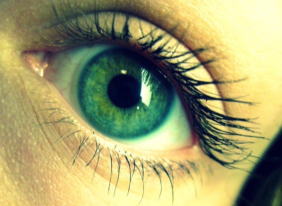
The Eyelid Margin Anatomy That Makes the Problem Possible
The eyelid margin is roughly 2 mm wide, and its architecture explains both how trichiasis develops and why certain treatments work. The critical landmark is the grey line - a faint visible demarcation separating the anterior lamella (skin, orbicularis muscle, and lash follicles) from the posterior lamella (tarsal plate, Meibomian glands, and conjunctiva). Every lash follicle sits at an angle within the anterior lamella, and that angle governs where the lash points. In a healthy eyelid, all follicles angle outward. Anything that distorts the tissue around a follicle - chronic inflammation, scarring, or structural lid disease - can tilt that angle inward.
The grey line is also the surgical landmark that makes lamellar splitting surgery possible. A precise incision along it separates the two lamellae cleanly, allowing the lash-bearing anterior strip to be repositioned without breaching either layer. Without that anatomical boundary, the separation would not be controllable.
Causes: The Variable That Predicts Treatment Difficulty
The most important distinction in trichiasis is not how many lashes are involved but whether the cause is cicatricial - meaning scar-driven - or non-cicatricial. This single variable predicts treatment difficulty and guides treatment selection more reliably than any other factor.
Non-cicatricial causes
- Chronic blepharitis - the most common cause in Western countries. Persistent lid-margin inflammation gradually thickens the tissue around follicles and shifts their growth angle inward. Because no permanent scarring has occurred, the follicular architecture responds reasonably to ablative treatments.
- Age-related lid laxity - structural changes over time can allow follicle angles to drift without underlying scar formation.
Cicatricial causes
- Trachoma - infection with Chlamydia trachomatis produces repeated conjunctival inflammation that scars the tarsal plate, deforms the lid margin, and rotates lashes inward. The WHO estimates trachoma is responsible for blindness or visual impairment in approximately 1.9 million people globally, making trachoma the world's leading infectious cause of preventable blindness.
- Stevens-Johnson syndrome - severe mucous membrane involvement leaves extensive conjunctival scarring throughout the posterior lamella.
- Ocular cicatricial pemphigoid - a progressive autoimmune blistering disease that continuously scars the conjunctiva over years.
- Herpes zoster ophthalmicus and herpes simplex conjunctivitis - viral infections leaving focal or diffuse lid-margin scarring after the acute episode resolves.
- Chemical burns - particularly alkali injuries, which cause deep structural change throughout the conjunctiva and lid margin.
- Prior eyelid surgery with poor healing - scar formation at the lid margin can permanently redirect follicle angles.
What Repeated Lash Contact Actually Does to the Cornea
The average person blinks roughly 15 to 20 times per minute. A single trichiatic lash therefore makes approximately 10,000 to 15,000 contact strokes against the corneal or conjunctival surface during every waking day. The damage accumulates in a recognizable sequence:
- Persistent irritation and reflex tearing. Foreign body sensation, photophobia, and excess tearing mark the earliest stage. Some patients with reduced corneal sensitivity from prior viral infection have surprisingly mild symptoms despite active lash contact - the absence of pain does not mean the cornea is safe.
- Corneal abrasion. Repeated contact strips superficial epithelial cells, creating punctate erosions visible under slit-lamp examination with fluorescein. These heal when the lash is removed but reopen as soon as it regrows.
- Corneal ulceration. Persistent epithelial breakdown allows bacteria to enter. At this stage, treatment requires more than lash removal alone, and systemic or topical antibiotic coverage becomes part of the management.
- Stromal scarring. Repeated ulceration reaching the corneal stroma leaves permanent opacity. Unlike epithelial wounds, stromal scars do not remodel fully, and each episode adds to cumulative opacity in the visual axis.
- Pannus formation and vascularization. In severe long-standing trichiasis, new blood vessels grow from the limbus inward across the corneal surface. This vascularization is irreversible and represents significant permanent vision loss - a progression thoroughly documented in trachoma-endemic regions where treatment remains inaccessible.
Lash stiffness, contact angle, and individual corneal sensitivity all influence how quickly a patient progresses through these stages. Some people tolerate lash contact for years with only mild discomfort; others develop significant corneal disease quickly. Neither outcome can be predicted from symptoms alone.
Temporary Relief: What Epilation Actually Is
Epilation - pulling the offending lash free with fine forceps at the slit lamp - provides immediate relief. For a patient with an acute corneal abrasion, it is the right first step. It is not a treatment. The follicle remains intact, and the lash regrows in the same misdirected direction within four to six weeks. Repeated epilation may also produce lashes that are coarser and stiffer, potentially more traumatic to the cornea over time.
Epilation has a legitimate role in the pathway: it controls symptoms while a more definitive plan is arranged, confirms which specific lashes are responsible for documented corneal changes, and is appropriate for patients not yet candidates for permanent treatment due to active lid-margin infection or inadequately controlled autoimmune disease.
The follicle that produced the inward-growing lash remains intact after epilation. The lash returns in the same wrong direction within four to six weeks - making repeated epilation a comfort measure, not a cure.
Ablative Treatments: Destroying the Follicle Without Surgery
All ablative treatments aim to damage the lash follicle permanently enough that it cannot produce another lash. Published success rates differ considerably by method, and the best choice depends on how many lashes are involved and whether the cause is cicatricial.
Electrolysis passes electrical current through a fine needle advanced to the follicle base, destroying germinal tissue. Published series report 48.9% of treated lids lash-free at six weeks and 67.8% success at a mean follow-up of 20 months; recurrence rates as high as 50% appear in other series. The technique requires precise needle placement and works best for focal involvement of roughly one to five lashes. Slight needle malpositioning leaves germinal tissue intact, allowing regrowth.
Cryoablation applies a double freeze-thaw cycle - reaching approximately -20 degrees Celsius or lower - directly to the lid margin. The two-cycle sequence is the operative mechanism: ice crystals form within cells during freezing and rupture cell membranes as they expand; the subsequent thaw compounds the osmotic injury. Rapidly dividing cells like follicle germinal epithelium are more vulnerable to this sequence than the slower-cycling connective tissue of the dermis and tarsal plate, which is why the technique can ablate follicles while sparing the structural integrity of the lid. One session achieves 34 to 56% lash clearance; two sessions reach 70 to 90%. Risks include lid notching from deeper dermal injury, permanent skin depigmentation (more visible and significant in darker skin tones), and potential reactivation of latent herpes zoster. Cryoablation treats a segment of lid margin simultaneously and is better suited to diffuse involvement than electrolysis.
Argon laser ablation directs a focused beam at individual follicles viewed through the slit lamp, producing targeted thermal destruction. Published data show 61.1% success after one session and 85.2% with up to three sessions. Reported complication rates vary across published series; some individual studies report hypopigmentation and lid notching each in roughly 8 to 12% of treated lids, while a 2025 systematic review and meta-analysis of 27 studies reported substantially lower pooled rates - patients should ask their provider about the specific complication profile associated with their technique and patient population.
Radiofrequency ablation delivers high-frequency energy through a fine probe to produce controlled thermal follicle destruction. It is used in some practices as an alternative to electrolysis for focal cases.
| Method | Best suited for | Published success | Key risk |
|---|---|---|---|
| Electrolysis | Focal (1-5 lashes) | ~68% at 20 months | Recurrence from incomplete ablation |
| Cryoablation | Diffuse, non-cicatricial | 70-90% after two sessions | Depigmentation, notching, zoster reactivation |
| Argon laser | Focal to moderate | 85.2% with three sessions | Hypopigmentation and notching (rates vary by series - confirm with provider) |
| Lamellar splitting | Diffuse, non-cicatricial | Durable; surgery-dependent | Lid contour change, surgical recovery |
| Mucous membrane graft | Cicatricial trichiasis | 55-92.6% in published series | Donor site morbidity, surgical complexity |
When Surgery Is the Answer
Ablative treatments work by destroying follicles one at a time. When involvement is diffuse across most of the lid margin, or when scarring has structurally distorted the lid itself, targeted follicle destruction cannot solve the underlying architectural problem. Surgery must address the cause directly.
Lamellar splitting for diffuse non-cicatricial trichiasis uses the grey line as a surgical hinge. An incision along the grey line separates the anterior lamella - carrying all the lash follicles - from the posterior lamella across the full eyelid width. The lash-bearing anterior strip is rotated outward and sutured in the everted position, redirecting every follicle away from the globe in a single procedure. Because the grey line provides a clean anatomical plane, the separation is precise and the structural integrity of both lamellae is maintained. The result is durable in diffuse non-cicatricial cases in a way that repeated focal ablation cannot achieve.
Mucous membrane grafting for cicatricial trichiasis addresses the root cause - scarred posterior lamella that is physically contracting and pulling follicles inward. A graft of buccal mucosa harvested from the inside of the cheek is inserted at the posterior lid margin to replace the scarred tissue. Expanding the posterior lamella releases the tethering that was misdirecting the lashes. Published series report success rates of 55 to 92.6%, with variation reflecting the severity of the underlying scarring and whether the causative disease is controlled.
Bilamellar tarsal rotation for trachomatous trichiasis incises the full-thickness eyelid across its width, rotates the posterior lamella to evert the lash line to a corrected position, and sutures the lid with lashes pointing away from the globe. The WHO endorses both bilamellar tarsal rotation and posterior lamellar tarsal rotation for trachomatous trichiasis; following randomized trial evidence showing lower postoperative trichiasis rates with the posterior lamellar technique, WHO guidance now recommends new surgeons be trained in posterior lamellar tarsal rotation, while surgeons already proficient in either approach may continue with the technique they know.
Choosing the Right Treatment
The framework clinicians use rests on four questions asked in sequence. First: is the lid margin in a normal position? If the entire lid rotates inward, the diagnosis is entropion and the treatment corrects that rotation - trichiasis-specific procedures will not hold. Second: how many lashes are involved? Focal cases suit electrolysis or laser; diffuse involvement points toward cryoablation or surgical correction of the lash line. Third: is the cause cicatricial? This is the question most patient-facing resources skip entirely. Ablative treatments have substantially lower success in cicatricial cases because the scarred tissue continues to distort new follicles after treated ones are gone. Fourth: has prior treatment already failed? Two rounds of ablation in a diffuse non-cicatricial case is reasonable grounds for moving to lamellar splitting rather than repeating a procedure with declining returns.
When to insist on an oculoplastic surgeon
- Diffuse trichiasis involving most of the lid margin
- Recurrence after two or more ablative treatment sessions
- Known cicatricial cause - trachoma, Stevens-Johnson syndrome, ocular cicatricial pemphigoid, or chemical burns
- Visible lid-margin distortion, notching, or structural irregularity
- Existing corneal scarring or pannus, indicating damage has progressed beyond irritation
- Suspected distichiasis rather than standard trichiasis
A general ophthalmologist can appropriately manage focal non-cicatricial trichiasis with electrolysis or laser. Once any of the above features are present, the case has moved into territory where lid architecture reconstruction - not follicle destruction - determines whether the result holds. That reconstruction requires oculoplastic training.
Frequently Asked Questions
Can trichiasis resolve on its own without treatment?
A misdirected lash may occasionally fall out and regrow at a better angle, but this cannot be counted on. Without correcting the follicle's growth angle, the lash returns in the same direction. If corneal contact is occurring, observation risks progressive corneal damage that accumulates silently before symptoms become severe.
Is trichiasis the same as an ingrown eyelash?
Trichiasis is the clinical term for misdirected lashes that contact the eye surface - which is what "ingrown eyelash" describes informally. The medical distinction matters because if the lid itself is rotated inward, the diagnosis shifts to entropion, and follicle removal alone will not correct the underlying lid rotation driving the contact.
Why does cryotherapy sometimes leave permanent white patches on the eyelid skin?
The freeze-thaw cycle destroys melanocytes - the pigment-producing cells in the skin - alongside the targeted follicle cells. In patients with darker skin tones, this depigmentation is more visible and is often permanent. It is worth discussing skin tone and pigmentation risk explicitly with the provider before choosing cryoablation over laser or electrolysis for focal cases where other options are equally available.
If argon laser works 85% of the time with three sessions, why would anyone need surgery?
The 85% figure reflects outcomes in non-cicatricial trichiasis. In cicatricial cases, all ablative treatments show substantially lower success because the scarred architecture continues to distort new follicles after treated ones are destroyed. Surgery that corrects the structural cause - mucous membrane grafting or tarsal rotation - produces more durable outcomes in those patients than additional ablation rounds.
Does trichiasis come back after surgery?
Surgical recurrence is possible, particularly when the underlying disease is active. Ocular cicatricial pemphigoid, for example, requires ongoing systemic immunosuppression; surgery without disease control carries a higher recurrence rate. In non-cicatricial diffuse trichiasis treated with lamellar splitting, recurrence is less common, but periodic follow-up is reasonable to catch individual follicles that may drift over time.
When should a patient seek an oculoplastic surgeon rather than returning to a general ophthalmologist?
A general ophthalmologist is well suited to focal non-cicatricial trichiasis managed with electrolysis or laser. The threshold for an oculoplastic referral is when the trichiasis is diffuse, recurrent after repeated ablation, cicatricial in origin, or accompanied by any lid-margin distortion - conditions where reconstructive eyelid surgery rather than follicle ablation is what the anatomy requires.
General information only, not medical advice. Individual anatomy and healing vary. See the disclaimer.