Most patients go into upper eyelid surgery thinking about skin. They know there is loose skin to remove, they expect to look rested, and they assume fat is simply something to be cleared away if present. What almost none of them have been told is that the upper eyelid contains two anatomically distinct fat compartments that age in opposite directions - and that treating them as a single undifferentiated target is one of the main reasons blepharoplasty results sometimes look hollowed and operated-on rather than natural. This guide explains what those compartments are, what age does to each of them separately, the risk posed by a structure at the outer lid that is not fat at all, and the specific questions that reveal whether a surgeon's approach to fat management is likely to serve you well over time.
Two Compartments, Not Three
The lower eyelid has three fat compartments: medial, central, and lateral. The upper eyelid has only two - the central preaponeurotic fat pad and the medial, or nasal, fat pad. There is no lateral fat compartment in the upper lid. This anatomical fact is well established, but its implications are almost never explained to patients, most of whom encounter blepharoplasty described as though the upper lid contains a single uniform mass of fat to be selectively reduced.
The two-compartment structure matters for surgery because the compartments differ in location, color, vascularity, and - most importantly - in how they respond to aging. A surgeon who approaches both compartments with the same removal philosophy, without accounting for their distinct behaviors, risks correcting one problem while creating another.
- Lower eyelid: three fat compartments (medial, central, lateral)
- Upper eyelid: two fat compartments only (central preaponeurotic, medial/nasal)
- No lateral fat pad exists in the upper lid
- Fullness at the outer corner of the upper lid is a structurally different problem than medial or central fullness - and requires a different assessment entirely
The Central Preaponeurotic Fat Pad
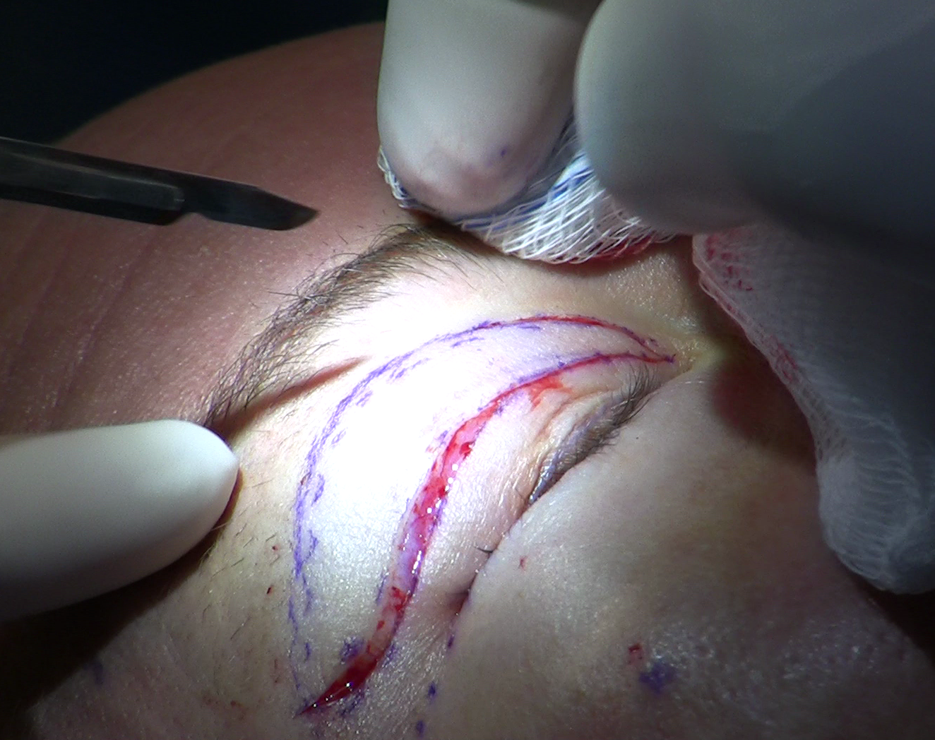
The central fat pad sits directly anterior to the levator aponeurosis - the fibrous sheet that physically lifts the eyelid when you open your eye. It is the first fat encountered when a surgeon opens the orbital septum during upper blepharoplasty. Because it lies immediately over the levator, careful dissection here matters not just cosmetically but functionally: injury to the levator aponeurosis produces ptosis, a drooped upper lid that requires separate repair.
The central fat pad is a distinctive yellow color. Surgeons use this coloration as an intraoperative confirmation that they are in the correct compartment. The color contrast with surrounding tissue is usually reliable under surgical lighting and helps distinguish it from the neighboring nasal fat, which is a different shade entirely.
What age does to this compartment is counterintuitive: the central preaponeurotic fat pad atrophies selectively with aging. It shrinks. Volume is progressively lost over decades, contributing to the sunken, hollow appearance that marks an older upper lid. For many patients presenting for blepharoplasty, the central compartment already has less fat than it should - which means indiscriminate removal from this area makes an existing deficiency worse.
The Medial (Nasal) Fat Pad
Anatomy and boundaries
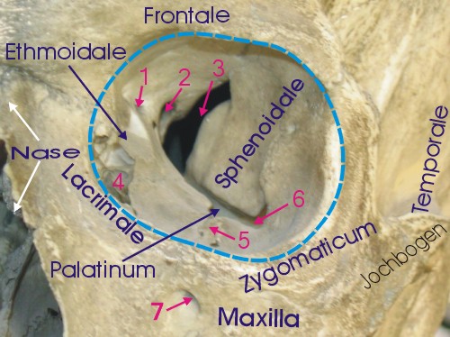
The nasal fat pad occupies the medial portion of the upper lid, separated from the central compartment by the trochlea of the superior oblique muscle and its fibrous attachments. The trochlea is a small fibrocartilaginous pulley structure through which the superior oblique tendon passes before it turns to reach its insertion on the eyeball. It serves as the anatomical boundary between the two compartments, and surgeons must positively identify it before entering the nasal compartment. Skipping that step carries real consequences.
Color distinguishes this pad immediately from its neighbor: nasal fat is white to pale rather than yellow. When a surgeon encounters white fat in the upper lid, it means either that they have crossed into the nasal compartment or that the trochlea boundary has not yet been confirmed. This color difference is a practical intraoperative safeguard.
Vascularity and the difficulty of working here
The nasal fat pad is the most vascularized of the two upper lid compartments. It has smaller lobules than the central fat, and the medial palpebral vessels run directly across it. This anatomy means that dissection in the nasal compartment produces more bleeding than dissection centrally, requires better hemostasis, and demands more careful visualization. It is partly why surgical literature identifies underresection of the medial fat as the most common reason for persistent upper eyelid fullness after blepharoplasty - the compartment is simply harder to work in confidently, and surgeons sometimes err toward caution at the cost of leaving more than the cosmetic result requires.
The Age Paradox: Opposite Trajectories in the Same Lid
This is the single most clinically important point that standard patient-facing blepharoplasty materials omit: the two fat compartments of the upper eyelid age in opposite directions. The central fat pad atrophies. The nasal fat pad does not - it is preserved or increases in volume with age. Same eyelid, same person, diverging paths.
CT volumetric studies have documented this differential, with the divergence typically becoming measurable around age 43. One proposed explanation is cellular: the nasal fat pad contains a higher concentration of neural-crest-derived progenitor cells than the central compartment. These cells may make nasal adipocytes more resistant to age-related atrophy, while the central fat - with lower concentrations of those protective cells - steadily shrinks.
The clinical implication is direct. In a patient in their 50s or 60s, the visible fullness or heaviness in the upper eyelid is almost always nasal fat. The central compartment has been atrophying for years already. A surgical approach that removes fat from both compartments without distinguishing between them will take from where volume is already deficient while possibly undertreating the structure actually responsible for the problem. That combination does not produce a refreshed result. It produces a lid that looks older after surgery than before it.
An older upper eyelid almost never has too much fat in both compartments at once. It has too much in the nasal compartment and too little in the central one - and surgery that ignores that difference will make the second problem worse.
The Lacrimal Gland: The Structure That Can Be Mistaken for Fat
Why the outer upper lid is a different problem
Because the upper eyelid has no lateral fat compartment, any fullness or prolapse at the outer corner of the upper lid is not fat until proven otherwise. The lacrimal gland - which produces the aqueous layer of the tear film - sits in a bony recess at the outer upper orbit. With age or as an individual variation, it can prolapse forward and appear as a soft fullness in the outer upper lid. When it does, it is visually indistinguishable from fat prolapse. Clinical palpation alone cannot reliably separate the two. CT imaging can confirm the distinction when there is any doubt.
Why removal is permanently damaging
Accidentally excising the lacrimal gland instead of repositioning it causes permanent aqueous tear deficiency. The gland does not regenerate. The result is severe, chronic dry eye disease - a debilitating condition affecting comfort, vision quality, and long-term ocular health. This is one of the most consequential errors in upper blepharoplasty. A prolapsed lacrimal gland should be identified, then repositioned back into its bony fossa, not removed. The distinction between a gland that needs repositioning and fat that might need conservative excision is exactly what imaging is for.
How the Field Moved from Aggressive Removal to Repositioning
Upper blepharoplasty has a history of aggressive fat removal. For decades the standard approach was to open the septum, identify whatever fat presented, and excise it. That logic - fat is the problem, removal is the solution - seems straightforward until you account for what age is doing to the compartments involved. Patients who had aggressive fat removal in their 40s returned in their 60s with lids that looked skeletal: a superior sulcus hollowed to the orbital rim, an appearance more aged than what brought them to surgery in the first place.
The field responded. Current oculoplastic and plastic surgery guidelines reflect a move toward conservative excision, deliberate preservation, and in many cases active repositioning of fat rather than its removal. The recognition that over-removal of fat - particularly central fat - creates superior sulcus hollowing drove this shift as much as improved anatomical understanding did.
| Approach | Fat philosophy | Hollowing risk | Best suited to |
|---|---|---|---|
| Aggressive removal | Excise all visible fat | Higher | Younger patients with true excess in both compartments |
| Conservative excision | Remove only what exceeds lid contour | Lower | Adults with moderate, compartment-specific fullness |
| Fat preservation | Avoid removal; address skin only | Minimal | Patients with early aging and central atrophy already present |
| Fat repositioning | Redirect nasal fat to refill central deficit | Addresses both simultaneously | Patients with nasal excess and central hollowing combined |
How Fat Repositioning Works in the Upper Lid
Repositioning solves the age-paradox problem surgically. When a patient has a prominent nasal fat pad alongside a depleted central compartment - increasingly the case after the mid-40s - the goal is not to simply remove the nasal excess. It is to redirect that fat into the central area that needs volume.
The technique involves carefully mobilizing the nasal fat pad without severing its vascular supply, then transposing it medially into the central sulcus where atrophy has created a deficit. The fat survives because its blood supply is preserved. It fills the hollow where central fat has been lost and simultaneously reduces the medial fullness that brought the patient to surgery - two problems addressed by one structure rather than creating a new one.
The outcomes data supports this approach. A study by Massry et al. examining nasal fat repositioning reported a 97% patient satisfaction rate at follow-up. A retrospective study using the validated Face-Q outcomes instrument found a 25-point improvement in upper eyelid satisfaction scores after volume-preservation blepharoplasty. A subbrow blepharoplasty study using fat transposition documented that the nasal fat pad grade (measuring the degree of visible protrusion) dropped by nearly a full grade while the central fat pad grade increased by more than a grade - demonstrating that the two deficits can be addressed simultaneously, with a 78% satisfaction rate.
Superior Sulcus Hollowing: What Over-Removal Produces
The superior sulcus is the groove between the upper eyelid and the orbital rim above it. In a naturally youthful eye, this area has gentle fullness from underlying fat. When that fat has been over-removed - either through aggressive primary blepharoplasty, cumulative procedures, or the progressive atrophy of central fat following any surgery - the sulcus becomes deeply hollow. The orbital rim becomes a visible hard ledge. The lid looks skeletal. Patients describe the appearance as "done," but not in a good way: the face looks older and more tired than it did before the surgery was performed.
Hollowing can also develop slowly after a surgery that looked acceptable initially. Fat continues to atrophy naturally with age, and a central compartment that was adequately preserved at 50 may look deficient at 65 as that process continues.
Correction options include:
- Hyaluronic acid filler injection into the superior sulcus - reversible, lower risk, requires maintenance, appropriate for mild to moderate hollowing
- Fat grafting using processed harvested fat - more permanent, requires multiple sessions, results vary with take rate
- Secondary surgical fat repositioning - a revision procedure to mobilize residual fat into the hollow, performed after adequate recovery time from the primary surgery
Surgeons also apply a minimum-distance rule requiring that enough skin remain between the lower border of the eyebrow and the upper lash margin after excision to allow complete eyelid closure - ask your surgeon for the specific threshold and how it applies to your anatomy. Violating it contributes to lagophthalmos (incomplete closure), which dries the cornea and creates its own category of long-term damage. Conservative skin excision is not timidity; it is a safeguard.
What to Ask a Surgeon About Fat Before You Agree to Surgery
Patients routinely review before-and-after photos, check board certifications, and ask about recovery time. Very few ask the questions that actually reveal how a surgeon thinks about fat. The decisions made about which compartments are opened, how much is removed, and whether any fat is repositioned will determine whether your result looks natural for decades or requires correction in five years. These questions are worth asking explicitly.
- Ask which compartments show excess or deficiency on your specific examination - a surgeon who has evaluated the central and nasal pads separately will give a compartment-by-compartment answer, not a general statement about "removing fat."
- Ask whether the outer upper lid fullness, if you have it, has been assessed for lacrimal gland involvement - and ask what the plan is if it turns out to be gland rather than fat.
- Ask whether the surgeon plans to reposition any fat or only remove it, and how they handle cases where the central compartment is already reduced.
- Ask what their policy is on the minimum skin bridge from brow to lash margin, and how much skin they plan to take from your lid specifically.
- Ask what the superior sulcus area should look like at five years and at ten years given your anatomy - this question forces a discussion about how ongoing aging will interact with what is removed.
The answers reveal depth of thinking about individual anatomy versus template-driven surgery. Watch for these distinctions in what you hear:
- Specific answer: distinguishes central from nasal fat, describes what was found in each compartment on your examination, includes criteria for repositioning versus removal, addresses the lacrimal gland question at the outer lid
- Vague answer: "we'll take out what's excess," "it depends on what we see," "we'll address the fat as needed" - with no compartment-level specificity when you push further
- Red flag: no mention of the trochlear boundary when discussing nasal fat; no protocol for assessing lacrimal gland involvement at the outer lid; dismissal of the central atrophy question
Frequently Asked Questions
Why does one part of my upper eyelid stay puffy as I age while another part looks hollow?
The two fat compartments in the upper lid age differently. The nasal (medial) fat pad resists atrophy and can remain prominent or increase in volume, while the central fat pad atrophies over time. CT volumetric studies place the onset of this divergence around age 43. The result - a lid with simultaneous excess in one area and deficiency in another - is the typical older upper eyelid, not a uniform fat accumulation problem.
Can my surgeon tell from looking whether outer upper lid fullness is fat or the lacrimal gland?
Not reliably, no. Visual inspection and clinical palpation alone cannot distinguish fat prolapse from lacrimal gland prolapse in many cases. When there is doubt, CT imaging should be used to confirm the structure before surgical decisions are made. A surgeon who proposes to address outer upper lid fullness without raising this question is taking a risk whose consequences - permanent dry eye from gland excision - cannot be undone.
If I had upper blepharoplasty years ago and my upper lid now looks hollow, what are my options?
Superior sulcus hollowing after prior surgery can be addressed by hyaluronic acid filler injection for a reversible and lower-risk correction, by fat grafting using processed harvested fat for a more permanent result, or by secondary surgical fat repositioning. Each approach has different risk profiles, longevity, and suitability depending on how much residual tissue is available and how severe the hollowing is. An oculoplastic surgeon with experience in secondary and revision cases is the appropriate specialist to evaluate your anatomy before recommending a route.
Why is nasal fat more often undertreated than central fat during surgery?
The nasal compartment is harder to work in. It is more vascularized than the central pad, bleeds more readily from vessels crossing it directly, and has a critical boundary structure - the trochlea - that must be confirmed before dissection. That complexity pushes surgeons toward caution in the nasal area, sometimes leaving more than the cosmetic result requires. Surgical literature identifies medial fat underresection as the most common reason for persistent upper eyelid fullness after blepharoplasty.
Does fat repositioning work for all patients, or only certain cases?
Repositioning is most useful when a patient has a prominent nasal fat pad alongside a deficient central compartment - the combination that becomes increasingly common after the mid-40s. It requires that the nasal pad have adequate volume to contribute and that there is a viable deficit in the central sulcus to receive it. Younger patients with uniform fat excess and preserved central volume may not need it at all and are better served by conservative excision. Your surgeon's preoperative assessment of each compartment separately is what determines whether repositioning applies to your anatomy.
How do I know if a surgeon's training covers these distinctions in depth?
Oculoplastic surgeons - whose specialty centers entirely on eyelid and orbital surgery - tend to have the deepest familiarity with compartment-specific anatomy, the lacrimal gland distinction, and repositioning techniques. That said, fellowship-trained plastic surgeons with a focused eyelid practice can also have this expertise. The consultation questions in this guide are designed to help you assess that depth regardless of a surgeon's formal specialty - the specificity of their answers about your anatomy is a more direct measure than credentials alone.
General information only, not medical advice. Individual anatomy and healing vary. See the disclaimer.