Squamous cell carcinoma of the eyelid is not the most common cancer of this region, but it is the one that keeps oculoplastic surgeons most alert. Basal cell carcinoma accounts for roughly 85 percent of malignant eyelid tumors while SCC accounts for approximately 5 percent - yet that smaller share carries a meaningfully higher risk of spreading to lymph nodes and beyond. For patients in South Florida, where published research documents SCC subtypes as more than 70 percent of all nonmelanoma skin cancers in some cohorts, understanding how this tumor forms, how it behaves differently from BCC, and why it demands a specialist team is not optional reading. It is practical self-defense.
What Eyelid SCC Is and Where It Fits in the Cancer Landscape
Squamous cells are the flat surface cells that line the skin. When chronic ultraviolet damage accumulates inside those cells over years, the DNA repair mechanisms fail and the cells begin to replicate abnormally. That process, repeated across thousands of cells on the lower eyelid, eventually produces a carcinoma - a cancer of epithelial origin.
The comparison with basal cell carcinoma is worth making directly. BCC is more common, grows more slowly, and almost never spreads to other parts of the body. SCC grows faster and metastasizes to regional lymph nodes or distant organs at rates reported between 1 and 21 percent depending on tumor characteristics - a range that underscores why the pathology report on an eyelid lesion matters so much. A low-risk SCC caught early carries an excellent prognosis. A high-risk SCC with nerve involvement caught late can track toward the orbit or skull base.
South Florida patients face a particular burden. Researchers studying nonmelanoma skin cancer in South Florida populations found that SCC subtypes now represent more than 70 percent of cases - a rate linked in part to decades of UV-B-blocking sunscreens that allowed cumulative UV-A damage to continue unremarked. Subtropical latitude, year-round outdoor activity, and the reflective surfaces of water and sand all amplify lifetime UV exposure in ways that make eyelid surveillance more urgent here than in most of the country.
Three Stages on the Road from Sun Damage to Invasive Cancer
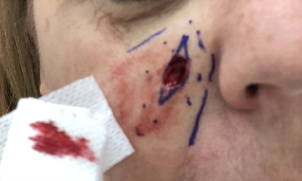
Understanding the stages between healthy skin and invasive SCC gives patients a clear picture of when intervention matters and what that intervention looks like at each point.
- Actinic keratosis. This is the earliest warning sign - a rough, scaly patch that forms on chronically sun-exposed skin. On the eyelid it typically appears on the lower lid margin or the medial canthal region. Two-thirds of eyelid SCCs arise from a previously identified actinic keratosis, which makes this lesion the single most important early signal. At this stage, the abnormal cells are confined to the surface layers of the epidermis. Cryotherapy, photodynamic therapy, or topical agents can eliminate the lesion before any invasive potential develops.
- SCC in situ - also called Bowen's disease or intraepidermal carcinoma. When abnormal cells fill the full thickness of the epidermis but have not yet broken through the basement membrane - the structural layer separating the skin surface from deeper tissue - the tumor is classified as in situ. It tends to look redder and more defined than an actinic keratosis, sometimes resembling an eczematous patch. Topical treatments including imiquimod and 5-fluorouracil can clear SCC in situ without surgery, though careful follow-up is needed to confirm the response.
- Invasive SCC. Once cells breach the basement membrane and extend into the dermis or deeper tissue, the tumor is invasive. Surgical excision is required, and the focus shifts to achieving clear margins while preserving as much eyelid anatomy as possible. This is the stage at which perineural spread, lymph node involvement, and distant metastasis become live considerations.
Each stage is a decision point. Recognizing an actinic keratosis on the eyelid and acting on it - rather than attributing it to dry skin or age - can prevent the progression to surgery altogether.
Who Is at Risk
Chronic UV exposure is the etiologic factor in approximately 90 percent of SCC cases. The lower eyelid and medial canthus bear the greatest cumulative UV dose of any eyelid surface because they lie in the direct path of both direct and reflected radiation. That said, UV load is not the only driver. Several independent factors raise the baseline risk substantially:
- Fair skin, light eyes, and a tendency to burn rather than tan - the skin types with the least innate UV protection
- A history of painful or blistering sunburns, which substantially increases SCC risk - a meta-analysis found that lifetime blistering sunburn history raises the odds of developing SCC by roughly 40 to 70 percent depending on frequency, with childhood exposures associated with even higher risk
- HPV infection, an independent risk factor separate from UV damage
- Long-term immunosuppressive therapy following organ transplantation - transplant recipients face a 65- to 250-fold increased SCC risk compared with the general population, and skin cancers are the most common malignancy in this group, accounting for an estimated 40 to 50 percent of all post-transplant malignancies, with SCC and BCC making up the large majority of those skin cancers
- Immunosuppression from other causes, including HIV infection and prolonged systemic corticosteroid use
- A prior diagnosis of SCC or multiple actinic keratoses, which signals a field of damaged skin rather than a single isolated abnormality
How Eyelid SCC Looks - and Why It Gets Misidentified
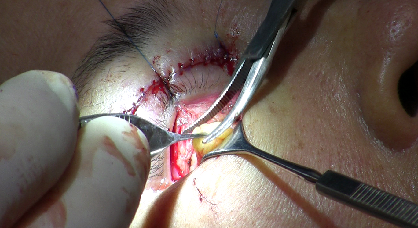
Eyelid SCC typically presents as an indurated, painless plaque or nodule on the lower lid or medial canthus. Unlike BCC, it generally lacks the pearly rolled border and surface blood vessels that most people associate with eyelid skin cancer. Instead, SCC tends to show hyperkeratosis - a rough, scaly, sometimes crusted surface - and it grows more rapidly than BCC, changing noticeably over weeks rather than months.
That absence of classic BCC features is part of what makes SCC easy to dismiss. Patients and even general practitioners sometimes attribute the lesion to a wart, a chalazion, an irritated cyst, or chronic blepharitis. The guiding principle is straightforward: any persistent eyelid lesion that does not resolve within a few weeks deserves a biopsy.
| Feature | Eyelid SCC | Eyelid BCC |
|---|---|---|
| Growth rate | Faster - weeks to months | Slower - months to years |
| Border appearance | Irregular, indurated, no pearly edge | Pearly, rolled border common |
| Surface texture | Rough, scaly, hyperkeratotic | Smooth or ulcerated |
| Risk of spread | Higher - regional nodes and beyond | Very low - local tissue only |
| Precursor lesion | Actinic keratosis, SCC in situ | No consistent precursor |
| Adjuvant treatment | Sometimes needed - radiation or node biopsy | Rarely needed |
Diagnosis: Biopsy, Staging, and What Perineural Invasion Means
A punch or shave biopsy of the suspicious area is the only reliable way to confirm the diagnosis. The pathology report will specify the degree of differentiation - well-differentiated tumors retain more normal cell structure and behave less aggressively; poorly differentiated tumors look markedly abnormal under the microscope and carry a higher risk of spread. Depth of invasion and margin status are also documented.
When imaging enters the picture
Most early eyelid SCCs do not require imaging before treatment. It becomes necessary when the tumor is large, when the patient reports symptoms suggesting orbital involvement such as double vision or restricted eye movement, or when perineural invasion is identified on biopsy. MRI with contrast is preferred in these cases because it outlines soft tissue planes and nerve anatomy better than CT; CT is added when bony orbital involvement is a concern.
Perineural invasion in plain terms
Perineural invasion is the process by which SCC cells grow along the sheath of a nerve, using it as a channel deeper into tissue. On the eyelid, this matters because the facial and trigeminal nerve branches supplying the lower lid and medial canthus connect, through progressively larger nerves, toward the orbital apex and skull base. A tumor that has invaded a nerve can track along that pathway toward structures surgery cannot safely reach.
Perineural invasion occurs in approximately 4.3 percent of periocular SCC cases, though some studies report higher rates depending on the population studied. When a pathologist identifies it, the clinical plan changes: imaging is ordered, adjuvant radiation is typically recommended, and lymph node assessment is considered. Some patients develop cranial nerve symptoms - numbness of the cheek, facial weakness, or changes in vision - before the nerve involvement is recognized. Any neurological symptom alongside an eyelid lesion warrants urgent evaluation.
Mohs Micrographic Surgery: Why Margin Control Is Different on the Eyelid
Standard excision removes the visible tumor with a margin of normal-appearing tissue, sends the specimen to an outside laboratory, and closes the wound - leaving the patient to wait days for the final margin result. Mohs micrographic surgery works differently. The surgeon removes the tumor and immediately processes horizontal sections of the entire peripheral and deep margin in an on-site laboratory, reading each layer while the patient waits. If cancer cells appear at any point on the margin, only that precise location is re-excised and re-examined, repeating until all margins are clear.
For eyelid SCC, this precision matters more than almost anywhere else on the face. The eyelid has almost no tissue to spare. Unnecessary removal in the wrong direction can compromise the lacrimal drainage system, stiffen the lid, or prevent full closure over the eye. Mohs surgery removes the minimum tissue required to achieve a clear margin.
Outcomes data support the approach. A five-year cure rate of 98.1 percent has been reported for SCC treated with Mohs surgery, with a periocular recurrence rate of approximately 5.9 percent. An Australian study specifically examining Mohs outcomes for periocular SCC found that patients with confirmed perineural invasion treated with Mohs surgery showed no recurrence at four to six years of follow-up.
At centers where this workflow functions as designed, a dermatologic surgeon performs and interprets the Mohs layers, and an oculoplastic surgeon performs the reconstruction in the same visit once margins are confirmed clear. This two-surgeon model is not a convenience - it is a clinical requirement. Reconstructing an eyelid before the final margin is confirmed risks closing tissue over residual cancer. It also means the reconstruction is performed by a surgeon who specializes specifically in eyelid anatomy, which is what drives the functional outcomes described below.
Eyelid Reconstruction: Function First, Appearance Second
The complexity of reconstruction depends on where the tumor sat, how much tissue was removed, and which anatomical structures - lid margin, tarsus, conjunctiva, lacrimal canaliculus - were affected. The oculoplastic surgeon's primary objective is always a lid that closes completely, protects the cornea, and allows tears to drain normally. Cosmetic symmetry follows from achieving those functional goals.
- Small defects can often be closed directly with fine sutures, preserving natural lid anatomy with minimal distortion
- Medium defects require local tissue flaps - the Tenzel semicircular flap recruits tissue from the lateral canthal area to bridge the gap without pulling the lid out of position
- Large defects, particularly those spanning more than half the lid, often require staged flaps from the opposing lid or cheek rotation flaps such as the Mustarde flap, which recruit larger amounts of skin while maintaining blood supply
- Medial canthal defects present additional complexity because the lacrimal canaliculi lie in this zone; reconstruction may require canalicular intubation to prevent post-operative chronic tearing
Published data show that oculoplastic reconstruction after periocular keratinocyte carcinoma achieves full eyelid function in more than 93 percent of patients and acceptable cosmetic appearance in more than 94 percent, with a complication rate below 3 percent. Those figures reflect what a surgeon with dedicated cancer reconstruction experience achieves - not a general average across all providers.
High-Risk Disease: Nerve Spread, Lymph Nodes, and When Radiation Is Added
Not every eyelid SCC follows the same management path. High-risk tumors require additional steps beyond surgery.
Sentinel lymph node biopsy maps and samples the first lymph node in the drainage pathway from the eyelid - typically in the parotid or submandibular region - to determine whether regional spread has occurred. Regional lymph node metastasis rates in high-risk periocular SCC range from roughly 1.3 to 24 percent depending on risk profile, which is wide enough to make individual tumor assessment essential. Finding cancer in the sentinel node changes the treatment plan to include complete lymph node dissection and radiation to the regional basin.
Adjuvant radiation is recommended in the following situations:
- Confirmed perineural invasion on pathology, even when surgical margins appear clear - nerve-tracked cells can extend beyond the visible surgical field
- Positive or close surgical margins that cannot be re-excised due to proximity to critical orbital structures
- Tumors near the orbital apex, where surgery cannot safely reach
- Regional lymph node involvement identified at sentinel node biopsy or clinical examination
Adjuvant radiation therapy is recommended when perineural invasion is confirmed on pathology or when margins cannot be fully cleared, particularly for tumors near the orbital apex.PubMed 28735439, Surgical Management of Periocular Cancers
Life After Treatment: Surveillance, Warning Signs, and Protecting What Remains
Clearing an eyelid SCC does not eliminate the risk of future disease. The same field of sun-damaged skin that produced the original tumor remains, and new actinic keratoses, new in-situ lesions, or a new invasive SCC can develop on the same or opposite eyelid. Structured follow-up with both the dermatologist and oculoplastic surgeon is standard practice, not an optional step taken only if something feels wrong.
Signs that warrant immediate evaluation rather than a wait-and-see approach
- Any new scaly, indurated, or crusted area on the treated or opposite eyelid that persists beyond two to three weeks
- A palpable lump in the cheek, jaw, or neck, which could represent lymph node enlargement
- Numbness or tingling of the cheek, forehead, or eyelid, which may suggest perineural spread
- Changes in vision, double vision, or restricted eye movement, which can indicate orbital involvement
- New or worsening tearing not present before surgery, which may indicate lacrimal disruption or recurrence near the medial canthus
Daily broad-spectrum sun protection applied to the periocular area - using products formulated for use near the eyes - along with wraparound UV-blocking sunglasses and avoidance of peak UV hours, reduces the risk of new actinic keratoses forming on remaining eyelid skin. Patients on immunosuppressive medications should discuss with their transplant or rheumatology team whether the regimen can be adjusted where medically safe, because reducing immune suppression where possible lowers ongoing skin cancer risk substantially.
A second opinion from an oculoplastic specialist is warranted when an eyelid lesion has been attributed to a benign cause for more than a month without improvement, when a biopsy has been performed but the treatment recommendation does not include Mohs surgery, or when any surgical plan involves removing eyelid tissue without a clear reconstruction plan from a surgeon who specializes in the eyelid.
Frequently Asked Questions
How is eyelid SCC different from basal cell carcinoma?
SCC grows faster and carries a meaningful risk of spreading to regional lymph nodes or distant organs - something BCC almost never does. SCC also tends to look rougher and scalier rather than having the pearly rolled border associated with BCC, and it more frequently requires additional steps such as lymph node assessment or adjuvant radiation alongside surgery. The two cancers share the same site and the same UV driver, but they behave differently enough that each requires its own clinical plan.
Can actinic keratosis on the eyelid be treated without surgery?
At the actinic keratosis stage, and at the SCC in situ stage, non-surgical treatments including topical imiquimod and 5-fluorouracil can clear abnormal cells before they become invasive. Once the tumor has broken through the basement membrane into deeper tissue, surgical excision is required. This is why acting on a persistent scaly eyelid lesion early matters - treating it at the actinic keratosis stage avoids the surgical pathway entirely.
What does it mean if my pathology report mentions perineural invasion?
Perineural invasion means SCC cells were found growing along the sheath of a nerve inside the removed tissue. This is the mechanism by which eyelid SCC can track from the skin surface toward the orbit and skull base. When it appears on the report, imaging is typically ordered to assess the extent of spread and adjuvant radiation is usually recommended - the surgical result alone is no longer sufficient, and follow-up becomes more intensive.
Why does eyelid SCC require two surgeons instead of one?
The Mohs surgeon removes the tumor and reads the tissue margins in real time until all margins are confirmed cancer-free; the oculoplastic surgeon then reconstructs the eyelid immediately after a clear result is in hand. Attempting reconstruction before margins are clear risks closing tissue over residual cancer. The oculoplastic surgeon also brings specific training in eyelid anatomy and lacrimal function that a general plastic or Mohs surgeon does not have, which is what produces functional results in more than 93 percent of cases.
Are organ transplant patients at significantly higher risk for eyelid SCC?
Yes. Organ transplant recipients on long-term immunosuppressive medications face a 65- to 250-fold increased SCC risk compared with the general population, and skin cancers are the most common malignancy in this group, accounting for approximately 40 to 50 percent of all post-transplant malignancies - with SCC and BCC comprising the large majority of those skin cancers. In South Florida, where UV exposure is year-round and intense, transplant patients need standing dermatology and oculoplastic surveillance rather than waiting for a visible or symptomatic lesion to prompt referral.
What does follow-up look like after Mohs surgery and reconstruction for eyelid SCC?
Follow-up typically includes scheduled visits with both the dermatologist and oculoplastic surgeon at intervals determined by the tumor's risk profile - lower-risk cases are seen less frequently, while high-risk cases with perineural invasion or lymph node involvement require more frequent clinical examination and sometimes imaging. Patients are counseled to self-examine and return immediately for any new lesion, nodal swelling, or neurological change rather than waiting for their next scheduled appointment, because early re-intervention carries far better outcomes than treating recurrence once it is established.
General information only, not medical advice. Individual anatomy and healing vary. See the disclaimer.