Contact lens wear is the single most common identifiable cause of acquired drooping eyelids in adults under fifty, accounting for nearly half of all such cases in one large surgical series. That figure surprises most patients, because ptosis is widely assumed to be a consequence of aging. In younger wearers, the cause is structural damage to a tendon - damage that accumulates quietly over an average of fifteen years before the lid drop becomes obvious. Understanding what breaks down, and why, clarifies why the fix requires surgery rather than a change in habits.
Why Long-Term Contact Lens Wear Can Droop the Eyelid
The eyelid margin descends not all at once but through a gradual progression that the body partially masks. In most cases the earliest visible change is a subtly elevated eyelid crease on one side, which appears before the lid margin itself drops noticeably. The frontalis - the broad forehead muscle - compensates by chronically elevating the brow, keeping lid opening functional for years. This is why the droop can look nearly normal in casual photographs: the brow is raised, the camera captures the compensated position, and both patient and onlookers underestimate the severity.
The underlying damage accumulates during ordinary daily activity. Thousands of blink cycles each day apply mechanical contact between the lid and lens surface. At any single moment the force is small. Applied repeatedly across years, the cumulative effect progressively weakens the aponeurotic attachment before the lid margin falls enough to draw attention.
This is not cosmetic aging. The anatomical endpoint - aponeurotic disinsertion or attenuation - belongs to the same category of change seen in age-related ptosis, but the driver is external and mechanical rather than the slow biochemical degradation of connective tissue with age. Recognizing the mechanical origin matters because it identifies an ongoing cause that stops only when the lens habit changes, and it points toward a surgical solution targeted specifically at the aponeurosis.
The Levator Aponeurosis: What the Tendon Does and Why It Is Vulnerable
The upper eyelid is raised primarily by the levator palpebrae superioris muscle, which originates at the back of the orbit and runs forward above the eye. The muscle itself does not attach directly to the lid margin. Instead, it transitions into a flat, sheet-like tendon called the levator aponeurosis, which then fans out and anchors to the front surface of the tarsal plate - the stiff cartilage-like structure that gives the eyelid its shape.
When the levator muscle contracts, it shortens and pulls the aponeurosis upward, which in turn pulls the tarsal plate and raises the lid margin. The system works efficiently when the aponeurosis is taut and firmly attached. The vulnerability comes from the aponeurosis being connective tissue rather than muscle. Connective tissue can be stretched, frayed, or pulled away from its attachment point by repeated mechanical force. Once stretched or disinserted, it does not recoil or heal back into position on its own.
The eyelid crease - the horizontal fold visible above the lid margin when the eye is open - forms where the aponeurosis sends fibers up through the orbicularis muscle to the skin. When the aponeurosis migrates superiorly because its lower attachment has loosened, it carries that crease upward with it. A crease sitting unusually high is one of the earliest visible signs that the tendon has begun to separate.
Two Mechanisms: Hard-Lens Removal Trauma vs. Soft-Lens Chronic Friction
The route from contact lens wear to ptosis differs significantly depending on lens type. Both pathways damage the same tendon, but they do so at different speeds and through different forces.
The Hard-Lens Removal Mechanism
A meta-analysis of hard contact lens wearers found an approximately seventeen-fold higher odds of blepharoptosis compared with non-wearers; subsequent research has also examined this elevated risk specifically in keratoconus patients who rely on RGP lenses long term. The mechanism is acute repetitive trauma. The standard hard-lens removal technique involves stretching the lateral eyelid outward with one finger while blinking to pop the lens free. Each repetition places a lateral traction force directly on the outer corner of the lid. Over thousands of removals across years, this traction progressively stretches the aponeurosis and eventually pulls it away from its tarsal attachment.
The Soft-Lens Chronic Friction Mechanism
Soft lens wearers face a slower, lower-force pathway. The edge of a soft lens exerts chronic low-level friction against the superior tarsal conjunctiva and the overlying orbicularis muscle every time the lid opens and closes - thousands of times per day. Over years, this sustained contact gradually loosens the aponeurotic attachment from below rather than pulling it from the side. The ptosis progresses more slowly than in hard-lens wearers but follows the same underlying aponeurotic pathology, as confirmed in a 2007 study published in Ophthalmology.
| Feature | Hard/RGP Lens Ptosis | Soft Lens Ptosis |
|---|---|---|
| Primary force | Acute lateral eyelid traction at removal | Chronic low-level edge friction |
| Relative risk | 17 times higher than non-wearers | Elevated; lower than RGP |
| Typical time to onset | Shorter; often under 15 years | Longer; typically 15 or more years |
| Mechanism site | Lateral canthal area, temporal aponeurosis | Superior tarsus and orbicularis |
| Underlying pathology | Aponeurotic disinsertion | Aponeurotic attenuation |
| Reversibility without surgery | None | None (partial swelling reduction only) |
How a Surgeon Identifies Contact-Lens Ptosis on Examination
The clinical examination of a patient with suspected contact lens-induced ptosis focuses on three measurements and one compensatory pattern that together confirm the aponeurotic origin.
Key Examination Findings
- Levator excursion above 10 mm, confirming the muscle itself is healthy and strong
- Eyelid crease sitting abnormally high or absent entirely on one side
- Lid margin lower than normal despite strong levator function
- Thin, almost translucent upper lid - because the aponeurosis no longer bulk-fills the space between muscle and tarsus
- Asymmetry between the two eyes, often correlating with which eye the patient manipulates more during lens removal
The elevated crease is the hallmark. In congenital ptosis, levator function is poor and the crease sits low or is absent. In aponeurotic ptosis - including the contact lens variety - the muscle works fine but its mechanical output is poorly transmitted because the tendon has migrated upward away from its anchor. The crease travels with it.
Frontalis Compensation
Patients with long-standing ptosis unconsciously recruit the frontalis - the broad forehead muscle - to lift the brow and pull the upper lid open by an indirect route. This compensation can make the ptosis appear milder than it is. During examination, a surgeon will manually stabilize the brow in a neutral position to reveal the true lid height. Patients who have compensated for years often present with chronic brow elevation, forehead tension, and headache that they have not connected to their eyelid at all.
Why Stopping Contact Lenses Will Not Reverse the Ptosis
The levator aponeurosis is connective tissue. It has no regenerative pathway comparable to muscle or epithelium. Once it has been stretched or pulled away from the tarsal plate, discontinuing lens wear stops further mechanical damage but cannot reattach the tendon to where it used to sit.
Some patients experience a small improvement in lid height after stopping lenses. This occurs because the chronic low-grade edema and irritation produced by the lens edge resolves, and with it a mild inflammatory component of the droop. That improvement addresses swelling, not structure. The underlying aponeurotic defect remains, and the ptosis will persist.
This is why the fifteen-year average wear duration matters. By the time the droop is obvious enough to bring a patient to a clinic, years of structural change have already accumulated. The good news is that the muscle remains entirely functional. Surgery works with an intact, healthy levator - it only needs to reconnect its tendon.
Upneeq Drops and Other Conservative Options: What They Can and Cannot Do
Upneeq (oxymetazoline hydrochloride ophthalmic solution 0.1%) is the only FDA-approved eye drop for acquired ptosis. It works by stimulating Muller's muscle, a smooth-muscle secondary elevator of the upper lid that runs alongside but independently of the levator. In phase 3 clinical trials, the drop raised lid height by approximately 1 mm on average, with individual results varying.
That is a meaningful lift for a patient with mild ptosis. For contact lens-induced ptosis, the drop serves a limited but specific role:
- As a temporary measure while a patient waits for surgery
- As a daily adjunct for patients with mild cases who choose not to pursue surgery
- As a diagnostic test - a good response to the drop confirms the ptosis is acquired aponeurotic type rather than a neurological problem
What Upneeq cannot do is address the disinserted aponeurosis. It bypasses the damaged pathway rather than repairing it, so the effect ends when the drop wears off. It does not slow the progression of the structural defect, and it offers no benefit to the crease abnormality or the asymmetry. For patients with moderate or significant ptosis, it typically produces insufficient lift on its own.
Conservative measures can manage symptoms temporarily, but only surgical reattachment of the aponeurosis corrects the underlying structural defect in contact lens-induced ptosis.
Levator Advancement Surgery: How the Operation Reattaches the Tendon
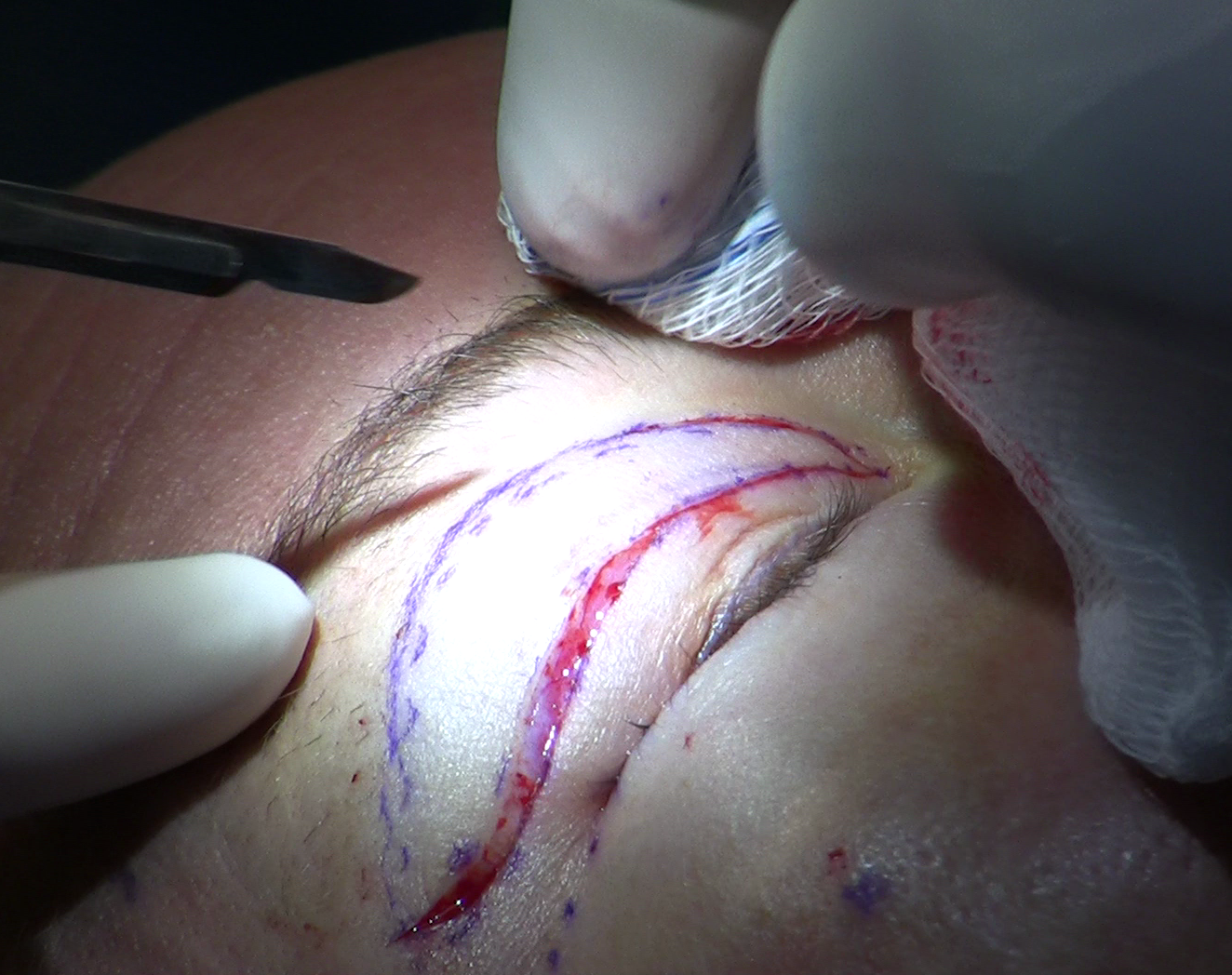
Levator advancement, also called aponeurotic repair, is the definitive treatment. The operation reconnects the disinserted or attenuated aponeurosis to its proper anchor point on the tarsus. Because the incision runs along the natural eyelid crease, it simultaneously allows removal of any excess upper lid skin, which is why the procedure is often combined with upper blepharoplasty when dermatochalasis is also present.
Step-by-Step Sequence
- The patient receives a local anesthetic injection into the upper eyelid; mild sedation is optional but the patient remains awake throughout.
- The surgeon marks and makes an incision precisely along the existing eyelid crease using a scalpel or fine cautery.
- Dissection proceeds through the orbicularis muscle to expose the orbital septum and then the aponeurosis beneath it.
- The disinserted or attenuated portion of the aponeurosis is identified and mobilized from surrounding tissue.
- The surgeon advances the aponeurosis downward and places temporary sutures attaching it to the anterior surface of the tarsus.
- The patient is asked to sit up and open the eyes so the surgeon can assess lid height and contour in a natural position.
- Suture tension is adjusted until the lid margin sits at the correct height and the crease appears symmetric with the opposite side.
- The skin incision is closed with fine sutures, reforming the eyelid crease at the proper position.
The awake adjustment step distinguishes this operation from most other procedures. Because lid height depends on the tension of the aponeurotic repair, intraoperative fine-tuning with a cooperative patient is more reliable than any measurement made while the eye is closed under general anesthesia.
Recovery, Return to Lenses, and Insurance Coverage
Healing Timeline
Swelling and bruising are expected for the first two weeks. The crease softens and settles into its final position over approximately six to eight weeks. During this period, strenuous activity and anything that raises blood pressure near the face should be limited. Lid height typically improves progressively as swelling resolves, and the final result is not fully assessable before the six-to-eight-week mark.
Returning to Contact Lens Wear
Most surgeons clear patients to resume contact lens wear after the crease has fully healed, generally around the six-to-eight-week point. Returning to wear is safe, but the original problem - eyelid manipulation during lens removal - has not changed. Aggressive removal techniques, particularly the lateral stretch-and-blink used for hard lenses, can gradually re-stress the repair over years. Many patients who reach this point choose to evaluate laser vision correction (LASIK or PRK) so that ongoing lens manipulation is no longer necessary. For patients with keratoconus who cannot pursue laser correction, careful technique with the minimum force needed for removal becomes a long-term priority.
Insurance and Functional Coverage
Ptosis surgery is covered as a functional procedure by many insurance plans when the droop is severe enough to interfere with vision. Coverage criteria are based on measurable thresholds - including the margin-to-reflex distance and the degree of superior visual field blocked by the lid - but those thresholds are set by individual insurers and can change. Patients should ask their surgeon to document the functional measurements accurately and then verify coverage criteria directly with their insurer before scheduling surgery.
Special Risk Groups
Keratoconus Patients
Keratoconus patients who rely on rigid scleral or gas-permeable lenses carry the highest identifiable risk. The larger diameter of a scleral lens requires more forceful and deliberate eyelid manipulation for both insertion and removal. These patients often begin wearing rigid lenses in adolescence or early adulthood and continue for decades. The seventeen-fold elevated risk established in RGP wearers applies with particular force here, because keratoconus patients typically cannot fall back on soft lenses or glasses as their vision deteriorates. Early, regular eyelid assessment should be part of their ongoing optometric and ophthalmologic care.
Decades-Long Wearers
Patients who have worn any contact lens type for more than ten years without a lid evaluation deserve a focused ptosis assessment even if they have not noticed a droop. The crease elevation that marks early aponeurotic change typically precedes a visible margin drop by several years, and because the frontalis compensates silently for a long time, a patient may pass casual self-inspection while structural change continues.
An examiner evaluating this group looks first at crease symmetry and position, then measures levator excursion to confirm the muscle is functioning normally, then manually neutralizes brow elevation to reveal true lid height. Finding a crease that sits higher on one side than the other - even without a measurable margin drop - is a reason to monitor closely. Identifying the problem before significant margin descent occurs generally means a less technically demanding repair when surgery becomes necessary.
Patients Scheduled for Blepharoplasty
Upper blepharoplasty and ptosis repair address different anatomical problems, and the presence of ptosis changes how much skin can safely be removed. The levator aponeurosis, when properly attached, sets the resting lid height. If the attachment is loose, the effective lid aperture is narrower than it should be, and some of the skin redundancy visible above the lid is a consequence of the low margin rather than true excess. Removing skin without correcting the underlying ptosis can deplete available skin margin and limit options for a future repair - or, if the lid height later shifts, leave an over-corrected appearance.
Surgeons planning a combined procedure account for ptosis correction first, then calculate skin excision relative to the corrected lid position. Any patient with a history of extended contact lens wear seeking blepharoplasty should ask specifically whether both conditions have been measured and whether the operative plan addresses them together. A combined correction in a single operation avoids a second procedure and produces a more predictable result.
Frequently Asked Questions
Can I develop ptosis from soft contact lenses, or only from hard lenses?
Both types carry risk. Hard lens wearers face a much higher relative risk because of the acute traction involved in removal, with studies showing approximately seventeen times the odds seen in non-wearers. Soft lens wearers develop ptosis through slower chronic friction rather than acute traction, but the resulting structural damage to the aponeurosis is the same type. A 2007 study published in Ophthalmology confirmed that young soft lens wearers - in that study, patients under thirty-five - develop aponeurotic ptosis on the same pathological basis as older adults with age-related lid droop.
How do I know whether my drooping lid is from contact lenses or just aging?
The two causes produce nearly identical anatomy and cannot be reliably distinguished by appearance alone. A surgeon will combine the clinical examination findings - elevated crease, strong levator function, thin lid - with a detailed history of contact lens use, wear duration, and removal technique. If contact lens wear has been long-term and the classic aponeurotic signs are present, the lens history is likely contributory even if other factors are also at play.
Will my ptosis come back after surgery if I keep wearing contact lenses?
Surgery repairs the aponeurosis but does not change the mechanical forces that damaged it. The risk of gradual recurrence is real for patients who return to aggressive removal technique over years. The earliest sign that a repair is being re-stressed is typically the same as the original presentation: the crease begins rising again on the affected side before the lid margin visibly drops. Noticing that change early - rather than waiting until the margin descends to its previous level - gives the best opportunity to address recurrence before it reaches the same severity as the original problem.
Is the surgery covered by insurance, or is it considered cosmetic?
Coverage depends on documented functional impairment measured by the margin-to-reflex distance and superior visual field testing. What determines approval in practice is thorough documentation: photographs taken with the brow in a neutral position, field-test results showing the degree of superior vision blocked, and a clinical note linking those measurements to the functional complaints the patient reports. Plans set their own thresholds and can change them, so pre-authorization verification with the insurer is necessary before scheduling. Patients who have been told a procedure is cosmetic should ask whether functional measurements were actually obtained before that determination was made.
Should I stop wearing contact lenses as soon as I notice a droop?
Stopping lens wear halts ongoing mechanical damage and allows any inflammatory swelling to resolve, but will not reattach the aponeurosis. During the six-to-eight-week pause before evaluation, using glasses instead, avoiding rubbing the lid, and letting any redness or irritation fully settle gives the most accurate picture of what structural droop remains once the inflammatory component is gone. If the lid is still down at that point, the problem is structural. The evaluation should include brow stabilization to reveal the true lid height, since frontalis compensation can make the remaining droop appear milder than it is.
Why does my eyelid crease look higher on the side where I droop?
A crease sitting higher on one side is an early structural signal worth acting on before the lid margin itself drops noticeably. A practical step is to photograph both eyes from the front with the brow consciously relaxed - not elevated - and compare crease positions and lid heights in the image. That photograph, combined with the contact lens history, gives an examining doctor a useful baseline for tracking whether the asymmetry is stable or progressing. Patients who notice this change before margin drop has occurred have identified the problem at its most correctable stage.
General information only, not medical advice. Individual anatomy and healing vary. See the disclaimer.