Thyroid eye disease forces the orbit to do something it was not built to handle: house more tissue than the fixed bony cavity can hold. The result is proptosis - eyes pushed visibly forward - along with pressure on the optic nerve, difficulty closing the eyelids, and in serious cases, rapid vision loss. Orbital decompression surgery is the mechanical answer to that crowding problem. It opens the walls of the orbit outward, giving displaced fat and swollen muscles room to expand rather than pressing forward against the cornea and optic nerve. This guide walks through exactly how that is done: which walls come down, what each removal adds to proptosis reduction and costs in double-vision risk, why the balanced two-wall approach has become the workhorse technique, what teprotumumab changed about who reaches the operating room, and what a realistic week-by-week recovery looks like.
What Proptosis Actually Does - and What Surgery Is Reversing
The orbit is a cone-shaped bony socket roughly 40 mm deep. Its volume is fixed. In thyroid eye disease, inflammation driven by IGF-1R and TSH receptor antibodies causes fat proliferation and extraocular muscle enlargement. When orbital volume increases but the walls cannot move, the contents push forward along the path of least resistance: out through the front opening, past the eyelids.
Three problems follow from sustained proptosis. First, the cornea - normally protected by the eyelids closing fully over it - becomes exposed when proptosis is severe enough that the lids no longer meet during sleep or even during a blink. Exposure keratopathy, corneal ulceration, and scarring are real consequences. Second, the enlarged extraocular muscles converge at the orbital apex, where the optic nerve exits through a narrow bony channel. Muscle pressure at the apex can compress the optic nerve directly - causing compressive optic neuropathy, a form of vision loss that can progress quickly. Third, mechanical displacement of the globe disrupts extraocular muscle alignment, producing double vision that is often worse on upgaze or lateral gaze.
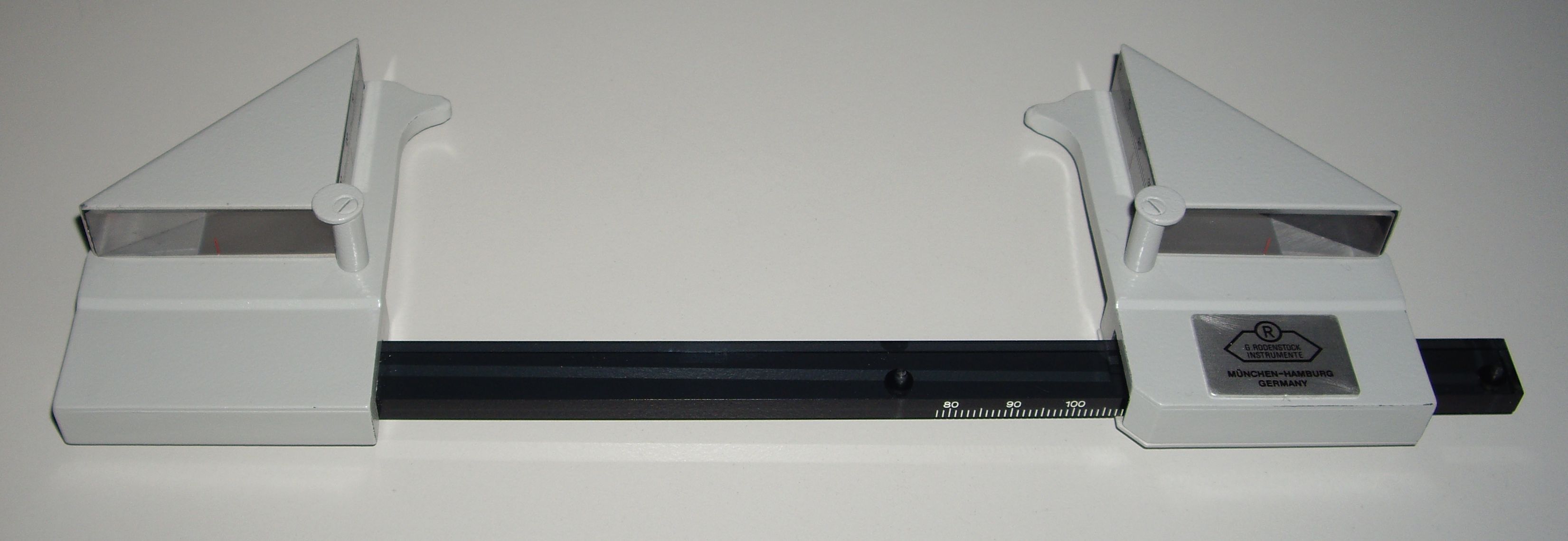
Orbital decompression surgery addresses all three by physically enlarging the bony orbit, letting the eye settle back toward its normal position. The goal - confirmed afterward by a Hertel exophthalmometer, which measures how far the globe protrudes beyond the lateral orbital rim - is to bring protrusion back to within about 2 mm of normal for the patient's population group.
The Four Walls of the Orbit - A Map of What Can Be Removed
The orbit is bounded by four walls, each with different thickness, neighboring structures, and surgical implications:
- Medial wall (lamina papyracea): The bone separating the orbit from the ethmoid air sinuses. Lamina papyracea means paper plate in Latin - an accurate description of a bone so thin it is nearly translucent. Because it already bows inward in some patients, it offers the least resistance when surgeons remove it, and doing so lets orbital fat herniate into the ethmoid cavity.
- Orbital floor: Below the globe, separated from the maxillary sinus by another thin bony shelf. Floor removal allows fat to drop into the sinus cavity. It contributes to proptosis reduction but carries a higher new diplopia risk than the lateral wall because the inferior rectus muscle - which controls downward gaze - runs directly above it.
- Lateral wall (greater wing of sphenoid): The thickest wall of the orbit, running from the orbital rim back into the temporal fossa. Surgeons must drill or burr it away, but removing it creates expansion room into the temporal fossa without disturbing any adjacent sinus. Critically, expanding into the temporal fossa does not displace the extraocular muscles as unpredictably as opening toward the ethmoid.
- Orbital roof: The thinnest wall above the globe, shared with the frontal sinus and the floor of the anterior cranial fossa. Roof decompression carries the highest risk of serious complications - including CSF leak and brain herniation - and is not performed in standard TED decompression cases.
Fat Decompression vs. Bone Decompression
Not all orbital decompression involves taking down walls. In patients with mild proptosis, or in those for whom bone work carries elevated risk, surgeons can remove orbital fat - both intraconal fat inside the muscle cone and extraconal fat outside it - without touching any wall.
Fat-only decompression produces roughly 2-4 mm of proptosis reduction on average. That may be sufficient when the presenting measurement is modestly elevated and the driver is fat proliferation rather than muscle enlargement. It also carries the lowest risk of new diplopia, because the muscles themselves are not mechanically displaced.
When fat removal is not enough - either because proptosis is severe, compressive optic neuropathy is present, or the muscles are massively enlarged - bone walls must come down. The two approaches can be combined in a single procedure: bone decompression sets the globe back by the amount dictated by wall removal, and fat removal then adds roughly 1-2 mm more on top of that while also preventing the hollowed appearance that sometimes follows when orbital volume drops sharply after bone work alone.
Wall by Wall - Proptosis Reduction and Diplopia Trade-offs
The choice of which wall or walls to remove is the central surgical decision in orbital decompression. Each combination produces different amounts of globe setback and different rates of new double vision. Because new diplopia - double vision that was not present before surgery - is the most consequential non-emergency complication, understanding these trade-offs is essential for anyone evaluating surgical options.
| Approach | Average proptosis reduction | New diplopia risk | Typical indication |
|---|---|---|---|
| Fat-only | 2-4 mm | Very low | Mild proptosis, cosmetic concern |
| Lateral wall alone | 2-3 mm | 0-6% | Mild-moderate, low diplopia tolerance |
| Medial wall alone | 2-3 mm | 10-35% | Less favored as a standalone approach |
| Balanced medial + lateral | 4-5.5 mm | 10-20% | Moderate-severe; workhorse approach |
| Deep lateral wall | Up to 11 mm | Lower than medial approaches | Severe proptosis, low diplopia risk priority |
| Three-wall (medial, lateral, floor) | Up to 7.5 mm | Higher; floor adds inferior rectus risk | Severe proptosis, refractory CON |
The logic behind balanced medial-plus-lateral decompression becomes clear from the medial-wall-alone diplopia figure. Removing only the medial wall produces new diplopia in 10-35% of cases - a wide range, mostly because fat herniating into the ethmoid pushes the medial rectus out of its normal alignment. Adding the lateral wall simultaneously provides expansion in the opposite direction, distributing the displacement more symmetrically and bringing the diplopia rate down to 10-20% while achieving 4-5.5 mm of reduction - roughly double what either wall alone would achieve. That combination of large reduction with controlled complication rate is why it has become the standard approach for most moderate-to-severe cases.
Deep Lateral Wall Decompression
Standard lateral wall removal stops near the orbital rim and anterior lateral wall. Deep lateral wall decompression takes drilling further back into the temporal fossa, well behind the rim and into the greater wing of the sphenoid. High-volume TED surgeons have increasingly favored this technique because it can achieve up to 11 mm of proptosis reduction - figures that previously required three-wall or four-wall approaches - while keeping the inferior orbital structures, including the inferior rectus muscle and the floor, entirely intact. That preservation keeps diplopia rates substantially lower than a three-wall approach at similar reduction levels, which is why the technique has displaced more aggressive multi-wall strategies as the preferred option when large corrections are needed.
Modern Incision Approaches - No Visible Scars
A concern patients frequently raise is scarring around an already affected area of the face. Modern surgical approaches for orbital decompression are designed to leave no visible marks:
- Swinging eyelid (lateral canthotomy plus lower fornix): The lateral wall is approached through a combination of a cut at the outer corner of the eye and an incision inside the lower lid. After surgery, the outer corner is re-sutured, and no skin scar remains.
- Upper eyelid skin crease: The lateral wall can also be accessed through the natural crease of the upper lid, where any scar is hidden in the fold.
- Transcaruncular: The medial wall is reached through a small incision at the caruncle - the pink tissue at the inner corner of the eye. It heals invisibly within the natural anatomy of the inner canthus.
- Transconjunctival: The orbital floor is accessed from inside the lower eyelid - in the conjunctiva - with no skin cut at all.
- Endoscopic trans-nasal: An ENT surgeon working alongside the orbital surgeon can remove the medial wall and floor entirely through the nose using endoscopes, with no skin or conjunctival incision whatsoever. This approach is available at centers where oculoplastic and sinus surgery teams collaborate but requires specialized equipment and training and is not universally offered. For patients to whom scar avoidance is a priority, it is worth asking about directly when evaluating surgical centers - teams at academic programs or dedicated TED clinics are the most likely setting.
Urgent vs. Elective - When Surgery Cannot Wait
The clearest emergency in thyroid eye disease is compressive optic neuropathy. When enlarged extraocular muscles compress the optic nerve at the orbital apex, the visual signal from the eye can be disrupted - sometimes rapidly. Signs that warrant urgent evaluation include:
- New or worsening blurred vision or color desaturation (colors appear washed out, particularly reds)
- A relative afferent pupillary defect - the pupil reacts more sluggishly to direct light than the other eye
- Visual field defects identified on formal testing
- Decreased contrast sensitivity
When compressive optic neuropathy is confirmed, decompression can and should be performed regardless of whether the disease is in the active or inactive phase. Waiting for stability would mean waiting while vision is being lost. For every other indication - severe proptosis, corneal exposure, orbital pain, disfigurement - elective timing rules apply, and the disease must be measurably stable for at least 3-6 months before the operation.
The rationale for requiring stability is mechanical rather than arbitrary. During the active inflammatory phase, orbital fat and muscle volumes are still changing. A decompression performed in an evolving orbit may produce excellent results on the table but shift unpredictably in the weeks afterward as swelling and fibrosis continue. Waiting for two or more stable Hertel measurements over that window ensures the anatomy being operated on is the anatomy the patient will end up with.
Tepezza First, Decompression Second - What Changed and What Did Not
Teprotumumab (Tepezza), an IGF-1R inhibitor given as a series of infusions, was approved in 2020 and significantly changed how many TED patients reach the operating room. Before effective medical therapy existed, historical data suggest roughly 85% of TED patients eventually required orbital decompression to manage proptosis. Studies of teprotumumab-treated patients show that only around 20-25% still need decompression after completing the infusion course - a substantial shift in the surgical landscape.
Teprotumumab and orbital decompression address the same disease but different phases of it: the drug acts on active inflammation, while surgery corrects the structural result that remains when inflammation has resolved or medical treatment has not fully reversed established proptosis.
The implications for patients are practical. Teprotumumab works best during the active inflammatory phase of TED - the window when antibody-driven fat proliferation and muscle swelling are still occurring. It has limited effect once the disease has entered its burned-out, fibrotic phase, because the structural changes at that point are no longer driven by ongoing inflammation. Patients who experience relapse after the infusion series, who have contraindications to the drug, or who present in inactive disease with established proptosis are clear surgical candidates.
The Rehabilitation Sequence - Why Order Is Not Optional
TED rehabilitation almost always involves more than one operation. Beyond decompression, many patients need strabismus surgery to correct persistent double vision and eyelid retraction repair to address the staring appearance and incomplete lid closure that often persist after proptosis is corrected. These operations must happen in a strict sequence, and understanding why each step depends on the one before it explains why no surgeon will skip ahead:
- Orbital decompression first. Moving the globe back into the socket changes the tension and alignment of the extraocular muscles. Any strabismus measurement taken before decompression reflects the pre-decompression eye position and will be inaccurate after the globe moves. Performing strabismus surgery on those pre-decompression numbers means correcting for a geometry that will change.
- Strabismus surgery second - typically 3-6 months after decompression. By this point, soft tissue remodeling is largely complete and the new globe position is stable. The strabismus surgeon measures the actual deviation in the new position and adjusts the muscles accordingly. Waiting the full interval - not rushing this step - matters because the final globe position may still be shifting at six weeks.
- Eyelid retraction repair last - after both globe position and muscle balance have stabilized. The position of the eyelids is directly influenced by both where the globe sits and the tension of the muscles running beneath the lid structures. Setting lid height before the globe and muscles have been corrected means measuring against anatomy that will change with the subsequent operations - almost certainly requiring further lid surgery to fix a result that was based on the wrong inputs.
The mechanical reason reversing this order causes problems is that each stage changes the input conditions for the next. A surgeon who attempts lid repair before muscle balance is established is setting a lid height against a moving target. The sequence exists because the anatomy is coupled - not because of arbitrary convention.
Recovery - Week by Week
Recovery from orbital decompression follows a consistent pattern, though its intensity varies with how many walls were removed and whether fat decompression was combined.
The First Week
Swelling and bruising are expected and peak at days 2-3. The bruising concentrates in the lower lids and cheeks when medial wall or floor work has been done, because blood tracks downward along tissue planes toward the face. The eyelids may swell enough to be temporarily difficult to open fully. Vision should be intact from the first day, though blurring from post-operative ointments and local swelling is common. The procedure is performed under general anesthesia as an outpatient - patients go home the same day - and typically takes 1-3 hours depending on the number of walls addressed.
Weeks Two Through Four
Swelling retreats progressively. Most patients are comfortable being seen in public by weeks two to three, though swelling is still visible. Many patients notice double vision during this period. This is usually edema-related - the swollen tissue is mechanically displacing the muscles - and it does not represent the final diplopia outcome. Cold compresses and head elevation help in the first days; after that, time is the primary factor.
The Six-Week Visit
By six weeks, most visible swelling has resolved. This is the first point at which surgeons assess whether new diplopia is present as a true surgical outcome rather than a temporary result of edema. If diplopia is confirmed by formal measurement at this visit, strabismus surgery will eventually be discussed - but no strabismus operation is considered until at least 3-6 months post-decompression, once the globe position has fully stabilized.
Three to Six Months - The Final Measurement
Soft tissue remodeling continues for months after bone work. The final Hertel measurement - the definitive confirmation of how much proptosis reduction was achieved - is typically taken at the 3-month visit and re-confirmed at six months before any further surgical planning begins. A result within about 2 mm of the population normal represents a successful decompression. If the residual proptosis is greater than that, additional surgery can be discussed - but only once two sequential measurements confirm the eye has stabilized. That stability is also what the strabismus surgeon waits for before making alignment measurements.
Frequently Asked Questions
How do surgeons decide how many walls to remove?
The decision is based on how much proptosis needs to be corrected, whether compressive optic neuropathy is present, whether the patient has pre-existing double vision, and how the patient weighs diplopia risk against the degree of globe setback achieved. A patient with modest protrusion and a cosmetic concern has different priorities than one with severe protrusion and optic nerve compression. High-volume TED surgeons discuss each approach's expected reduction range and diplopia rate before surgery so the patient understands the specific trade-offs for their situation - not a generic one.
Will I definitely have double vision after surgery?
Not necessarily, but it is a real possibility depending on which walls are removed. Fat-only and lateral wall decompression carry the lowest rates - roughly 0-6% for new diplopia. Balanced medial-plus-lateral carries a 10-20% rate, and adding the orbital floor increases this further. Early postoperative double vision is common and usually resolves as swelling clears; persistent diplopia assessed at the six-week visit is a different and separate finding, one that would be addressed with strabismus surgery later in the rehabilitation sequence if it remains stable.
Can orbital decompression be done if teprotumumab did not work?
Yes. Teprotumumab is effective primarily during the active inflammatory phase of TED; once the disease has burned out and proptosis remains, the drug cannot reverse fibrotic tissue changes. Patients who saw no response, partial response, or relapse after completing the infusion series are routine surgical candidates. The surgery addresses the structural result of the disease rather than the inflammatory process, so it works regardless of prior medical treatment history.
Why can the eyelids not be repaired at the same time as decompression?
Because orbital decompression changes the position of the globe and the tension on the extraocular muscles - both of which influence eyelid height. Eyelid retraction repair involves setting the lid at a specific height relative to the cornea. If that height is set before the globe has been moved back and the muscles have been rebalanced, the lid will shift when subsequent operations alter the underlying mechanics - requiring further lid surgery to correct a result that was based on the wrong anatomy. The strict sequence exists because the three systems are coupled, not independent.
What does the Hertel measurement after surgery actually mean?
The Hertel exophthalmometer rests on the lateral orbital rims and measures how many millimeters the cornea protrudes beyond that reference point. TED patients often present with values of 23-28 mm; decompression aims to bring that number to within about 2 mm of the normal range for the patient's population group. The final measurement at three to six months confirms whether the globe has settled to that target and whether the eye position is stable enough for any subsequent strabismus surgery to be meaningfully planned. Two stable sequential measurements - not just one reading - give the strabismus surgeon a reliable target to work from.
Is endoscopic trans-nasal decompression widely available?
No. Endoscopic approaches to the medial wall and orbital floor require collaboration between an oculoplastic surgeon and an ENT or skull base surgeon with sinus endoscopy expertise, as well as specific equipment. Centers that perform high volumes of TED surgery - particularly those affiliated with academic medical programs or dedicated thyroid eye disease clinics - are the most likely setting to find teams with that combined training. When evaluating surgical options, asking specifically whether endoscopic trans-nasal decompression is offered, and how many procedures the team has performed that way, is a reasonable and practical question. The American Society of Ophthalmic Plastic and Reconstructive Surgery (ASOPRS) maintains a surgeon directory that can help identify oculoplastic specialists who focus on TED rehabilitation.
General information only, not medical advice. Individual anatomy and healing vary. See the disclaimer.